• 文献检索
  • 文档翻译
  • 深度研究
  • 学术资讯
  • Suppr Zotero 插件Zotero 插件
  • 邀请有礼
  • 套餐&价格
  • 历史记录
应用&插件
Suppr Zotero 插件Zotero 插件浏览器插件Mac 客户端Windows 客户端微信小程序
定价
高级版会员购买积分包购买API积分包
服务
文献检索文档翻译深度研究API 文档MCP 服务
关于我们
关于 Suppr公司介绍联系我们用户协议隐私条款
关注我们

Suppr 超能文献

核心技术专利:CN118964589B侵权必究
粤ICP备2023148730 号-1Suppr @ 2026

文献检索

告别复杂PubMed语法,用中文像聊天一样搜索,搜遍4000万医学文献。AI智能推荐,让科研检索更轻松。

立即免费搜索

文件翻译

保留排版,准确专业,支持PDF/Word/PPT等文件格式,支持 12+语言互译。

免费翻译文档

深度研究

AI帮你快速写综述,25分钟生成高质量综述,智能提取关键信息,辅助科研写作。

立即免费体验

通过计算机视觉与基于融合的联合迁移学习在多模态医学图像中的集成,实现乳腺癌的增强诊断。

Enhanced breast cancer diagnosis through integration of computer vision with fusion based joint transfer learning using multi modality medical images.

机构信息

Department of Computing Technologies, School of Computing, SRM Institute of Science and Technology, Kaatankulathur, Chennai, 603203, India.

Department of Computer Science and Engineering, Periyar Maniammai Institute of Science & Technology, Vallam, Thanjavur, 613403, India.

出版信息

Sci Rep. 2024 Nov 17;14(1):28376. doi: 10.1038/s41598-024-79363-6.

DOI:10.1038/s41598-024-79363-6
PMID:39551870
原文链接:https://pmc.ncbi.nlm.nih.gov/articles/PMC11570594/
Abstract

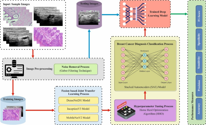
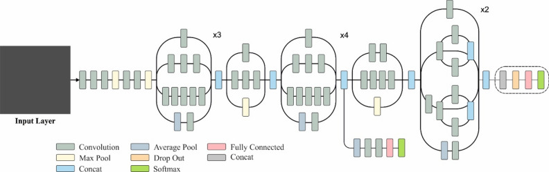
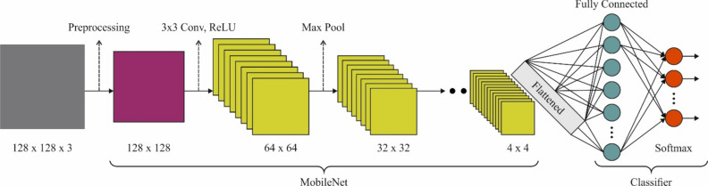
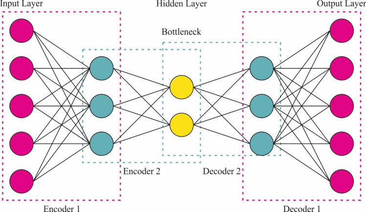
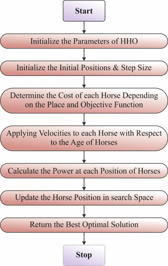
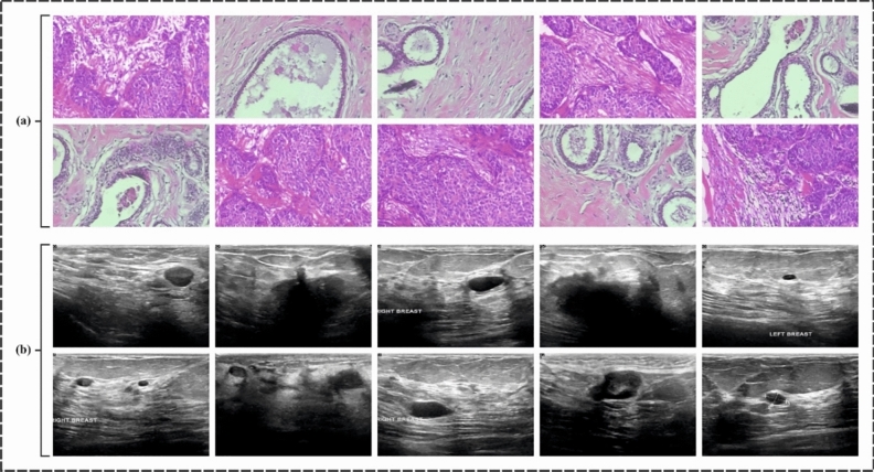
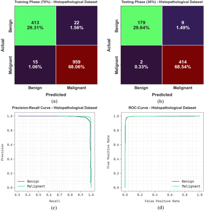
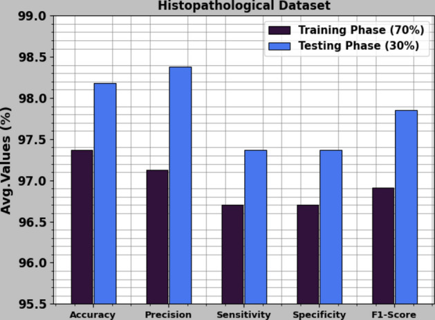
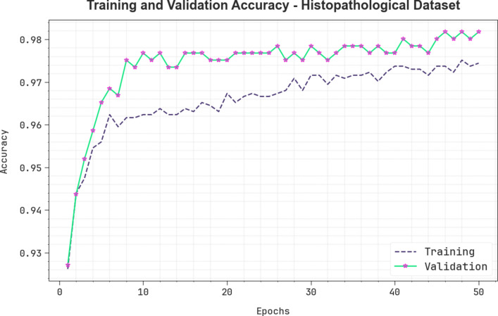
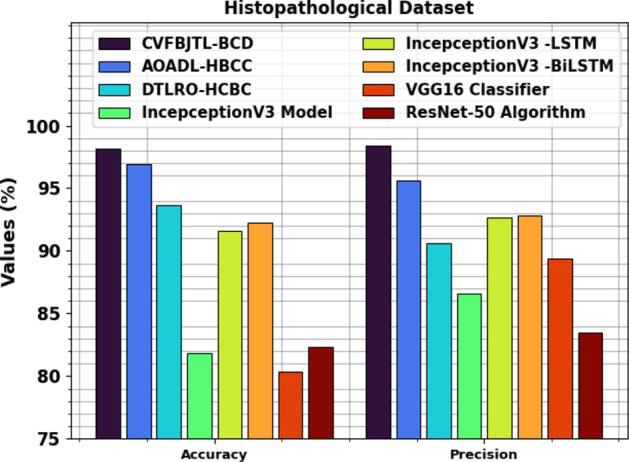
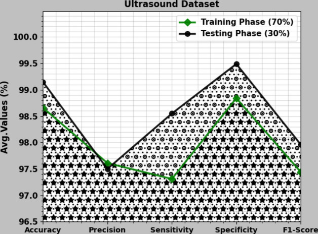
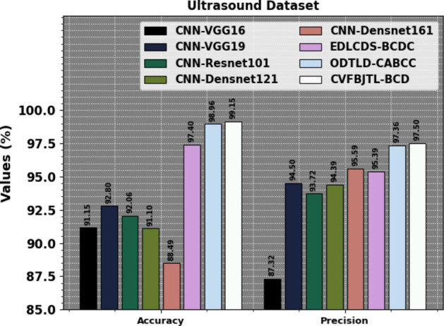
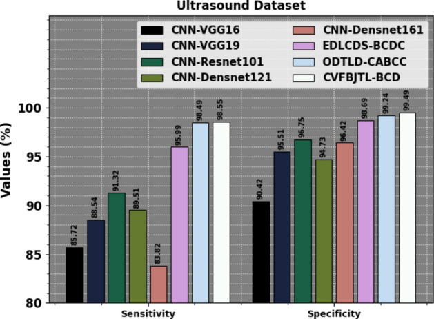
Breast cancer (BC) is a type of cancer which progresses and spreads from breast tissues and gradually exceeds the entire body; this kind of cancer originates in both sexes. Prompt recognition of this disorder is most significant in this phase, and it is measured by providing patients with the essential treatment so their efficient lifetime can be protected. Scientists and researchers in numerous studies have initiated techniques to identify tumours in early phases. Still, misperception in classifying skeptical lesions can be due to poor image excellence and dissimilar breast density. BC is a primary health concern, requiring constant initial detection and improvement in analysis. BC analysis has made major progress recently with combining multi-modal image modalities. These studies deliver an overview of the segmentation, classification, or grading of numerous cancer types, including BC, by employing conventional machine learning (ML) models over hand-engineered features. Therefore, this study uses multi-modality medical imaging to propose a Computer Vision with Fusion Joint Transfer Learning for Breast Cancer Diagnosis (CVFBJTL-BCD) technique. The presented CVFBJTL-BCD technique utilizes feature fusion and DL models to effectively detect and identify BC diagnoses. The CVFBJTL-BCD technique primarily employs the Gabor filtering (GF) technique for noise removal. Next, the CVFBJTL-BCD technique uses a fusion-based joint transfer learning (TL) process comprising three models, namely DenseNet201, InceptionV3, and MobileNetV2. The stacked autoencoders (SAE) model is implemented to classify BC diagnosis. Finally, the horse herd optimization algorithm (HHOA) model is utilized to select parameters involved in the SAE method optimally. To demonstrate the improved results of the CVFBJTL-BCD methodology, a comprehensive series of experimentations are performed on two benchmark datasets. The comparative analysis of the CVFBJTL-BCD technique portrayed a superior accuracy value of 98.18% and 99.15% over existing methods under Histopathological and Ultrasound datasets.

摘要

乳腺癌(BC)是一种从乳腺组织发展和扩散并逐渐超过全身的癌症;这种癌症起源于两性。在这个阶段,及时发现这种疾病是最重要的,通过为患者提供必要的治疗来保护他们的有效寿命来衡量。许多研究的科学家和研究人员已经开始使用技术来识别早期阶段的肿瘤。然而,由于图像质量差和乳房密度不同,对可疑病变的分类可能存在误解。BC 是一个主要的健康关注点,需要不断进行初步检测和分析改进。BC 分析最近在结合多模态图像模式方面取得了重大进展。这些研究通过在手工制作的特征上使用传统的机器学习(ML)模型,提供了乳腺癌等多种癌症类型的分割、分类或分级概述。因此,本研究使用多模态医学成像,提出了一种用于乳腺癌诊断的计算机视觉融合联合转移学习技术(CVFBJTL-BCD)。所提出的 CVFBJTL-BCD 技术利用特征融合和深度学习模型来有效检测和识别 BC 诊断。CVFBJTL-BCD 技术主要采用 Gabor 滤波(GF)技术去除噪声。接下来,CVFBJTL-BCD 技术使用基于融合的联合转移学习(TL)过程,该过程包括三个模型,即 DenseNet201、InceptionV3 和 MobileNetV2。使用堆叠自动编码器(SAE)模型对 BC 诊断进行分类。最后,利用马群优化算法(HHOA)模型对 SAE 方法中涉及的参数进行最优选择。为了证明 CVFBJTL-BCD 方法的改进结果,在两个基准数据集上进行了一系列全面的实验。CVFBJTL-BCD 技术的比较分析在组织病理学和超声数据集上分别表现出 98.18%和 99.15%的卓越准确性值,优于现有方法。

https://cdn.ncbi.nlm.nih.gov/pmc/blobs/b63e/11570594/cbbe98bfa6f0/41598_2024_79363_Fig22_HTML.jpg
https://cdn.ncbi.nlm.nih.gov/pmc/blobs/b63e/11570594/9747d96383ea/41598_2024_79363_Fig1_HTML.jpg
https://cdn.ncbi.nlm.nih.gov/pmc/blobs/b63e/11570594/f2dd3091382b/41598_2024_79363_Fig2_HTML.jpg
https://cdn.ncbi.nlm.nih.gov/pmc/blobs/b63e/11570594/fdefa5f97453/41598_2024_79363_Fig3_HTML.jpg
https://cdn.ncbi.nlm.nih.gov/pmc/blobs/b63e/11570594/ef86858a4d25/41598_2024_79363_Fig4_HTML.jpg
https://cdn.ncbi.nlm.nih.gov/pmc/blobs/b63e/11570594/9204538d7e51/41598_2024_79363_Fig5_HTML.jpg
https://cdn.ncbi.nlm.nih.gov/pmc/blobs/b63e/11570594/d9b586d961a0/41598_2024_79363_Fig6_HTML.jpg
https://cdn.ncbi.nlm.nih.gov/pmc/blobs/b63e/11570594/c0e10e86379d/41598_2024_79363_Fig7_HTML.jpg
https://cdn.ncbi.nlm.nih.gov/pmc/blobs/b63e/11570594/c34714bae267/41598_2024_79363_Fig8_HTML.jpg
https://cdn.ncbi.nlm.nih.gov/pmc/blobs/b63e/11570594/8f4a5493513c/41598_2024_79363_Fig9_HTML.jpg
https://cdn.ncbi.nlm.nih.gov/pmc/blobs/b63e/11570594/b35662c25c06/41598_2024_79363_Fig10_HTML.jpg
https://cdn.ncbi.nlm.nih.gov/pmc/blobs/b63e/11570594/d0a6079a54ce/41598_2024_79363_Fig11_HTML.jpg
https://cdn.ncbi.nlm.nih.gov/pmc/blobs/b63e/11570594/ace09d1e1a8b/41598_2024_79363_Fig12_HTML.jpg
https://cdn.ncbi.nlm.nih.gov/pmc/blobs/b63e/11570594/96e768cc883f/41598_2024_79363_Fig13_HTML.jpg
https://cdn.ncbi.nlm.nih.gov/pmc/blobs/b63e/11570594/0f22d4d229c1/41598_2024_79363_Fig14_HTML.jpg
https://cdn.ncbi.nlm.nih.gov/pmc/blobs/b63e/11570594/da8bc054fac5/41598_2024_79363_Fig15_HTML.jpg
https://cdn.ncbi.nlm.nih.gov/pmc/blobs/b63e/11570594/a81492dc701a/41598_2024_79363_Fig16_HTML.jpg
https://cdn.ncbi.nlm.nih.gov/pmc/blobs/b63e/11570594/5ede859c6109/41598_2024_79363_Fig17_HTML.jpg
https://cdn.ncbi.nlm.nih.gov/pmc/blobs/b63e/11570594/74cb158870bf/41598_2024_79363_Fig18_HTML.jpg
https://cdn.ncbi.nlm.nih.gov/pmc/blobs/b63e/11570594/b9dd7bc4bbb5/41598_2024_79363_Fig19_HTML.jpg
https://cdn.ncbi.nlm.nih.gov/pmc/blobs/b63e/11570594/db1fdfe954cb/41598_2024_79363_Fig20_HTML.jpg
https://cdn.ncbi.nlm.nih.gov/pmc/blobs/b63e/11570594/04d5b891d99a/41598_2024_79363_Fig21_HTML.jpg
https://cdn.ncbi.nlm.nih.gov/pmc/blobs/b63e/11570594/cbbe98bfa6f0/41598_2024_79363_Fig22_HTML.jpg

相似文献

1
Enhanced breast cancer diagnosis through integration of computer vision with fusion based joint transfer learning using multi modality medical images.通过计算机视觉与基于融合的联合迁移学习在多模态医学图像中的集成,实现乳腺癌的增强诊断。
Sci Rep. 2024 Nov 17;14(1):28376. doi: 10.1038/s41598-024-79363-6.
2
ViT-MAENB7: An innovative breast cancer diagnosis model from 3D mammograms using advanced segmentation and classification process.基于先进分割和分类流程的 3D 乳腺 X 线摄影的乳腺癌诊断新模型:ViT-MAENB7。
Comput Methods Programs Biomed. 2024 Dec;257:108373. doi: 10.1016/j.cmpb.2024.108373. Epub 2024 Aug 23.
3
Hybrid machine learning-based breast cancer segmentation framework using ultrasound images with optimal weighted features.基于混合机器学习的乳腺癌超声图像分割框架,使用最优加权特征。
Cell Biochem Funct. 2024 Jun;42(4):e4054. doi: 10.1002/cbf.4054.
4
Advanced feature learning and classification of microscopic breast abnormalities using a robust deep transfer learning technique.使用稳健的深度迁移学习技术对微观乳腺异常进行高级特征学习和分类。
Microsc Res Tech. 2024 Aug;87(8):1862-1888. doi: 10.1002/jemt.24557. Epub 2024 Mar 30.
5
[Breast cancer lesion segmentation based on co-learning feature fusion and Transformer].基于协同学习特征融合与Transformer的乳腺癌病灶分割
Sheng Wu Yi Xue Gong Cheng Xue Za Zhi. 2024 Apr 25;41(2):237-245. doi: 10.7507/1001-5515.202306063.
6
Infrared thermal images using PCSAN-Net-DBOA: An approach of breast cancer classification.基于 PCSAN-Net-DBOA 的红外热图像:一种乳腺癌分类方法。
Microsc Res Tech. 2024 Aug;87(8):1742-1752. doi: 10.1002/jemt.24550. Epub 2024 Mar 19.
7
Enhanced multimodal medical image fusion via modified DWT with arithmetic optimization algorithm.基于改进的 DWT 与算术优化算法的增强型多模态医学图像融合。
Sci Rep. 2024 Aug 20;14(1):19261. doi: 10.1038/s41598-024-69997-x.
8
Towards laryngeal cancer diagnosis using Dandelion Optimizer Algorithm with ensemble learning on biomedical throat region images.基于生物医学喉部图像的 Dandelion Optimizer 算法集成学习进行喉癌诊断。
Sci Rep. 2024 Aug 24;14(1):19713. doi: 10.1038/s41598-024-70525-0.
9
Fusing global context with multiscale context for enhanced breast cancer classification.融合全局上下文和多尺度上下文以提高乳腺癌分类。
Sci Rep. 2024 Nov 9;14(1):27358. doi: 10.1038/s41598-024-78363-w.
10
[Research progress of breast pathology image diagnosis based on deep learning].基于深度学习的乳腺病理图像诊断研究进展
Sheng Wu Yi Xue Gong Cheng Xue Za Zhi. 2024 Oct 25;41(5):1072-1077. doi: 10.7507/1001-5515.202311061.

引用本文的文献

1
Multimodal integration strategies for clinical application in oncology.肿瘤学临床应用中的多模态整合策略
Front Pharmacol. 2025 Aug 20;16:1609079. doi: 10.3389/fphar.2025.1609079. eCollection 2025.

本文引用的文献

1
A Dual-Branch Cross-Modality-Attention Network for Thyroid Nodule Diagnosis Based on Ultrasound Images and Contrast-Enhanced Ultrasound Videos.一种基于超声图像和超声造影视频的双分支跨模态注意力甲状腺结节诊断网络。
IEEE J Biomed Health Inform. 2025 Feb;29(2):1269-1282. doi: 10.1109/JBHI.2024.3472609. Epub 2025 Feb 10.
2
SAMA: A Self-and-Mutual Attention Network for Accurate Recurrence Prediction of Non-Small Cell Lung Cancer Using Genetic and CT Data.SAMA:一种利用基因和CT数据进行非小细胞肺癌准确复发预测的自注意力与互注意力网络。
IEEE J Biomed Health Inform. 2025 May;29(5):3220-3233. doi: 10.1109/JBHI.2024.3471194. Epub 2025 May 6.
3
An explainable AI-based blood cell classification using optimized convolutional neural network.
一种基于可解释人工智能的血细胞分类方法,采用优化的卷积神经网络。
J Pathol Inform. 2024 Jul 2;15:100389. doi: 10.1016/j.jpi.2024.100389. eCollection 2024 Dec.
4
Investigating the detection of breast cancer with deep transfer learning using ResNet18 and ResNet34.使用 ResNet18 和 ResNet34 的深度迁移学习检测乳腺癌。
Biomed Phys Eng Express. 2024 Apr 18;10(3). doi: 10.1088/2057-1976/ad3cdf.
5
A twin convolutional neural network with hybrid binary optimizer for multimodal breast cancer digital image classification.一种具有混合二进制优化器的双卷积神经网络,用于多模态乳腺癌数字图像分类。
Sci Rep. 2024 Jan 6;14(1):692. doi: 10.1038/s41598-024-51329-8.
6
FP-CNN: Fuzzy pooling-based convolutional neural network for lung ultrasound image classification with explainable AI.FP-CNN:基于模糊池化的卷积神经网络,用于具有可解释人工智能的肺部超声图像分类。
Comput Biol Med. 2023 Oct;165:107407. doi: 10.1016/j.compbiomed.2023.107407. Epub 2023 Sep 1.
7
Hyperparameter Optimizer with Deep Learning-Based Decision-Support Systems for Histopathological Breast Cancer Diagnosis.基于深度学习的决策支持系统的超参数优化器用于乳腺癌组织病理学诊断
Cancers (Basel). 2023 Jan 31;15(3):885. doi: 10.3390/cancers15030885.
8
Incorporating a Novel Dual Transfer Learning Approach for Medical Images.将一种新的双重迁移学习方法应用于医学图像。
Sensors (Basel). 2023 Jan 4;23(2):570. doi: 10.3390/s23020570.
9
DR-IIXRN : Detection Algorithm of Diabetic Retinopathy Based on Deep Ensemble Learning and Attention Mechanism.DR-IIXRN:基于深度集成学习和注意力机制的糖尿病视网膜病变检测算法
Front Neuroinform. 2021 Dec 24;15:778552. doi: 10.3389/fninf.2021.778552. eCollection 2021.
10
Deep learning for diabetic retinopathy detection and classification based on fundus images: A review.基于眼底图像的糖尿病视网膜病变检测和分类的深度学习:综述。
Comput Biol Med. 2021 Aug;135:104599. doi: 10.1016/j.compbiomed.2021.104599. Epub 2021 Jun 25.