Chen Junxiao, Wang Ruixue, Dong Wei, He Hua, Wang Shiyong
Department of Information, Third Affiliated Hospital of Naval Medical University, No. 225 Changhai Road, Yangpu District, 200438, Shanghai, China.
Department of Neurosurgery, Third Affiliated Hospital of Naval Medical University, No. 225 Changhai Road, Yangpu District, 200438, Shanghai, China.
BMC Med Imaging. 2025 Jan 7;25(1):9. doi: 10.1186/s12880-025-01550-2.
To develop an end-to-end convolutional neural network model for analyzing hematoxylin and eosin(H&E)-stained histological images, enhancing the performance and efficiency of nuclear segmentation and classification within the digital pathology workflow.
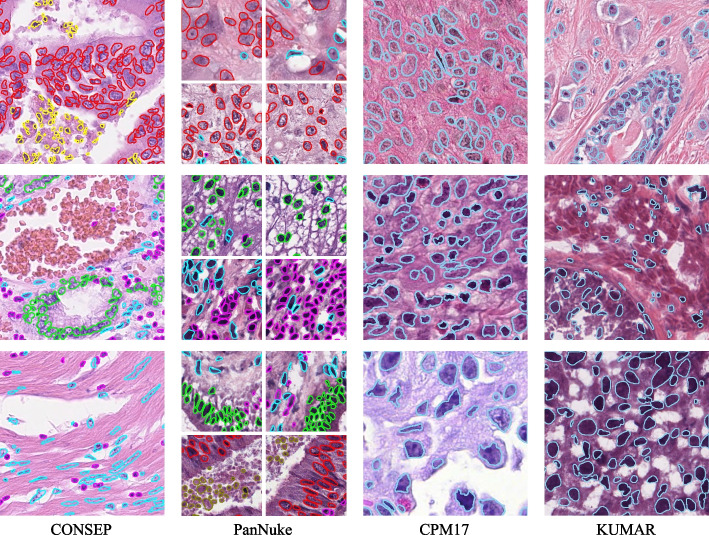
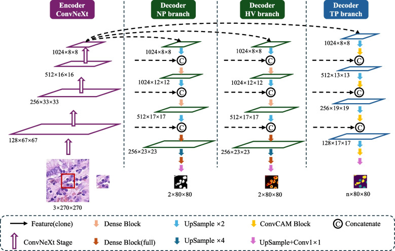
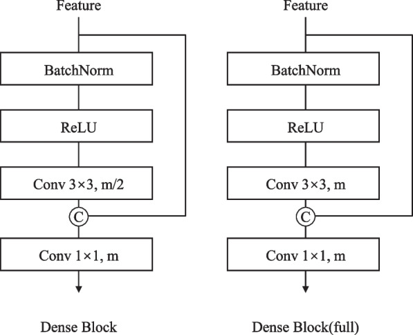
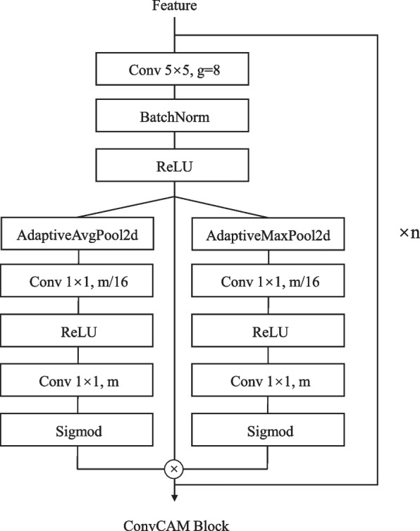
We propose a dual-mechanism feature pyramid fusion technique that integrates nuclear segmentation and classification tasks to construct the HistoNeXt network model. HistoNeXt utilizes an encoder-decoder architecture, where the encoder, based on the advanced ConvNeXt convolutional framework, efficiently and accurately extracts multi-level abstract features from tissue images. These features are subsequently shared with the decoder. The decoder employs a dual-mechanism architecture: The first branch of the mechanism splits into two parallel paths for nuclear segmentation, producing nuclear pixel (NP) and horizontal and vertical distance (HV) predictions, while the second mechanism branch focuses on type prediction (TP). The NP and HV branches leverage densely connected blocks to facilitate layer-by-layer feature transmission and reuse, while the TP branch employs channel attention to adaptively focus on critical features. Comprehensive data augmentation including morphology-preserving geometric transformations and adaptive H&E channel adjustments was applied. To address class imbalance, type-aware sampling was applied. The model was evaluated on public tissue image datasets including CONSEP, PanNuke, CPM17, and KUMAR. The performance in nuclear segmentation was evaluated using the Dice Similarity Coefficient (DICE), the Aggregated Jaccard Index (AJI) and Panoptic Quality (PQ), and the classification performance was evaluated using F1 scores and category-specific F1 scores. In addition, computational complexity, measured in Giga Floating Point Operations Per Second (GFLOPS), was used as an indicator of resource consumption.
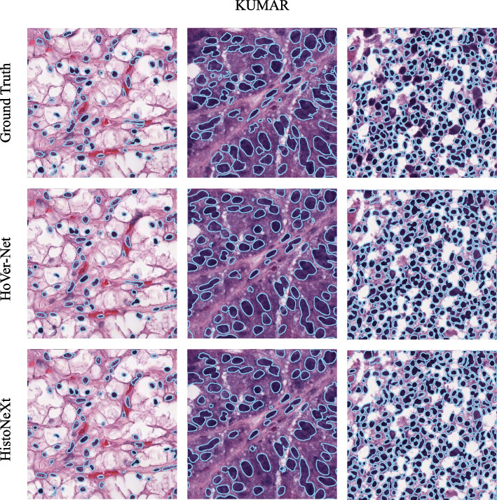
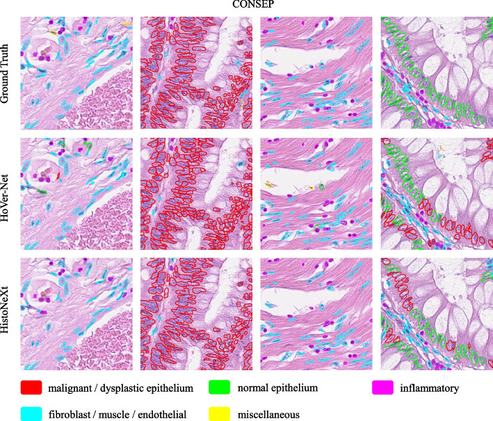
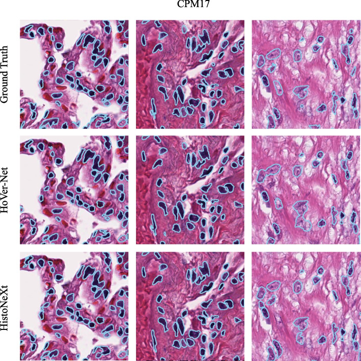
HistoNeXt demonstrated competitive performance across multiple datasets: achieving a DICE score of 0.874, an AJI of 0.722, and a PQ of 0.689 on the CPM17 dataset; a DICE score of 0.826, an AJI of 0.625, and a PQ of 0.565 on KUMAR; and performance comparable to Transformer-based models, such as CellViT-SAM-H, on PanNuke, with a binary PQ of 0.6794, a multi-class PQ of 0.4940, and an overall F1 score of 0.82. On the CONSEP dataset, it achieved a DICE score of 0.843, an AJI of 0.592, a PQ of 0.532, and an overall classification F1 score of 0.773. Specific F1 scores for various cell types were as follows: 0.653 for malignant or dysplastic epithelial cells, 0.516 for normal epithelial cells, 0.659 for inflammatory cells, and 0.587 for spindle cells. The tiny model's complexity was 33.7 GFLOPS.
By integrating novel convolutional technology and employing a pyramid fusion of dual-mechanism characteristics, HistoNeXt enhances both the precision and efficiency of nuclear segmentation and classification. Its low computational complexity makes the model well suited for local deployment in resource-constrained environments, thereby supporting a broad spectrum of clinical and research applications. This represents a significant advance in the application of convolutional neural networks in digital pathology analysis.
开发一种端到端卷积神经网络模型,用于分析苏木精和伊红(H&E)染色的组织学图像,提高数字病理学工作流程中细胞核分割和分类的性能与效率。
我们提出了一种双机制特征金字塔融合技术,将细胞核分割和分类任务整合起来构建HistoNeXt网络模型。HistoNeXt采用编码器-解码器架构,其中编码器基于先进的ConvNeXt卷积框架,从组织图像中高效准确地提取多级抽象特征。这些特征随后与解码器共享。解码器采用双机制架构:该机制的第一分支分为两条并行路径进行细胞核分割,生成细胞核像素(NP)以及水平和垂直距离(HV)预测,而第二个机制分支专注于类型预测(TP)。NP和HV分支利用密集连接块促进逐层特征传递和重用,而TP分支采用通道注意力自适应地聚焦于关键特征。应用了包括保持形态的几何变换和自适应H&E通道调整在内的综合数据增强方法。为解决类别不平衡问题,采用了类型感知采样。该模型在包括CONSEP、PanNuke、CPM17和KUMAR在内的公共组织图像数据集上进行评估。细胞核分割性能使用骰子相似系数(DICE)、聚合雅卡尔指数(AJI)和全景质量(PQ)进行评估,分类性能使用F1分数和特定类别F1分数进行评估。此外,以每秒千兆浮点运算(GFLOPS)衡量的计算复杂度被用作资源消耗的指标。
HistoNeXt在多个数据集上表现出具有竞争力的性能:在CPM17数据集上,DICE分数为0.874,AJI为0.722,PQ为0.689;在KUMAR数据集上,DICE分数为0.826,AJI为0.625,PQ为0.565;在PanNuke数据集上,其性能与基于Transformer的模型(如CellViT-SAM-H)相当,二元PQ为0.6794,多类别PQ为0.4940,总体F1分数为0.82。在CONSEP数据集上,它实现了DICE分数为0.843,AJI为 0.592,PQ为0.532,总体分类F1分数为0.773。各种细胞类型的特定F1分数如下:恶性或发育异常上皮细胞为0.653,正常上皮细胞为0.516,炎症细胞为0.659,梭形细胞为0.587。小型模型的复杂度为33.7 GFLOPS。
通过整合新颖的卷积技术并采用双机制特征的金字塔融合,HistoNeXt提高了细胞核分割和分类的精度与效率。其低计算复杂度使该模型非常适合在资源受限环境中进行本地部署,从而支持广泛的临床和研究应用。这代表了卷积神经网络在数字病理学分析应用中的重大进展。