Schmitz-Moormann P, Himmelmann G W, Brandes J W, Fölsch U R, Lorenz-Meyer H, Malchow H, Soehendra L N, Wienbeck M
Gut. 1985 Apr;26(4):406-14. doi: 10.1136/gut.26.4.406.
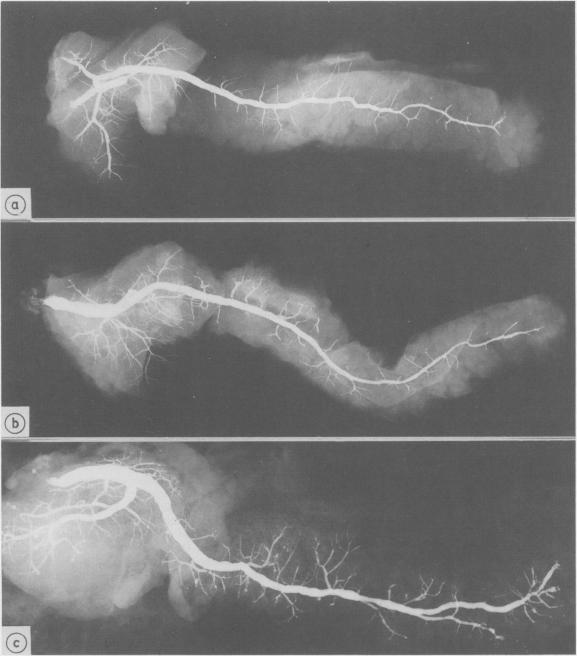
A postmortem study by ductography and histology was performed on 69 human pancreata with no clinical or histological signs of chronic pancreatitis. The ductograms, supplemented by five postmortem ductograms of chronic pancreatitis, were independently evaluated by six clinicians, skilled in ERCP; the degree of alteration was estimated by simple rating, forced choice rating, and by determination of the grade of chronic pancreatitis, Histologically, the amount of intraductal epithelial proliferation, periductal, intralobular and perilobular fibrosis, intraductal protein plugs, and fat necrosis was determined by semiquantitative methods. The six ductographical evaluations significantly differed in the level of their data, but corresponded in the range of distribution. All evaluations were correct regarding judgement of ductograms from patients with chronic pancreatitis. Ductograms of patients without chronic pancreatitis, however, were also frequently classified as chronic pancreatitis; overall 81% (minimal 37%, moderate 33%, severe 11%). This high level of false positive diagnosis indicates the frequency of pancreatitis like lesions in the main duct and its side branches in patients without chronic pancreatitis. Ductal lesions were significantly correlated with perilobular fibrosis. This finding favours the assumption, that in the non-inflamed pancreas, perilobular fibrosis plays a key-role in the development of ductal alterations, as in chronic pancreatitis. Perilobular fibrosis may result from intralobular inflammation caused by age-dependent intraductal epithelial hyperplasia.
对69例无慢性胰腺炎临床或组织学征象的人体胰腺进行了胰管造影和组织学的尸检研究。6名擅长内镜逆行胰胆管造影(ERCP)的临床医生对这些胰管造影图(并补充了5例慢性胰腺炎的尸检胰管造影图)进行了独立评估;通过简单评分、强制选择评分以及确定慢性胰腺炎的分级来评估改变程度。组织学上,通过半定量方法确定导管内上皮增生、导管周围、小叶内和小叶周围纤维化、导管内蛋白栓子以及脂肪坏死的量。六种胰管造影评估的数据水平有显著差异,但分布范围一致。对于慢性胰腺炎患者的胰管造影判断,所有评估都是正确的。然而,无慢性胰腺炎患者的胰管造影图也经常被归类为慢性胰腺炎;总体为81%(轻度37%,中度33%,重度11%)。这种高假阳性诊断率表明无慢性胰腺炎患者的主胰管及其分支中类似胰腺炎病变的发生率。导管病变与小叶周围纤维化显著相关。这一发现支持了这样一种假设,即在非炎症性胰腺中,小叶周围纤维化在导管改变的发展中起关键作用,如同在慢性胰腺炎中一样。小叶周围纤维化可能由年龄依赖性导管内上皮增生引起的小叶内炎症导致。