Svanekjaer Laura, Larsen Jeppe K P, Frederiksen Peter H, Linde Louise, Gregers Emilie, Udesen Nanna L J, Helgestad Ole K, Banke Ann, Jensen Lisette O, Lassen Jens F, Povlsen Amalie L, Schmidt Henrik, Møller Jacob E, Ravn Hanne B
Department of Cardiothoracic Anaesthesiology, Odense University Hospital, J.B. Winsløwsvej 4, 5000, Odense, Denmark.
Department of Cardiology, Odense University Hospital, Odense, Denmark.
Intensive Care Med Exp. 2025 Sep 1;13(1):91. doi: 10.1186/s40635-025-00802-3.
Low systolic blood pressure (SBP) is a key criterion for diagnosing cardiogenic shock (CS) caused by a reduction in stroke volume and cardiac output (CO). The temporal interaction between changes in pressure and flow has not been well described in the development of CS. In a large animal model, we assessed the temporal relationships of SBP, CO, and blood flow in the carotid artery during induction of CS.
Fifteen adult Danish landrace pigs (median weight 71 kg) underwent CS induction by stepwise injection of polyvinyl alcohol microspheres into the left main coronary artery every 3 min to induce microvascular obstruction. After each injection, CO, SBP, and mixed venous saturation (SvO) were recorded simultaneously from a ventricle sheath in the carotid artery and a pulmonary artery catheter in the right internal jugular vein. A Doppler flow probe measured blood flow in the left carotid artery. CS was defined as a ≥ 50% reduction in CO or SvO from baseline, or absolute SvO < 30%.
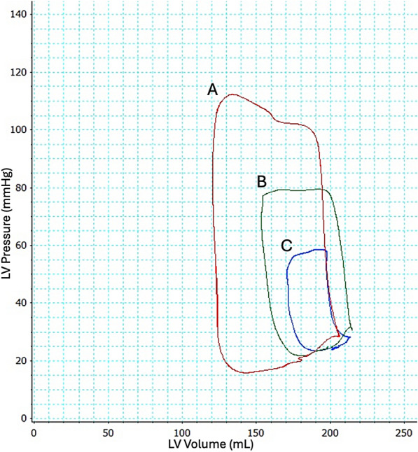
CS occurred after a mean of 8 (range 5 to 19) boluses of microspheres. SBP declined from 99 (± 15) mmHg to 74 (± 6) mmHg, equal to 74 (± 13)% of the baseline value. CO was reduced to 5.8 (± 0.7) L/min to 2.2 (± 1.3) L/min, equal to 38 (± 23)% and SvO from 63 (± 7)% to 37 (± 7)%, equal to 60 (± 13)% of baseline values. The decrease in CO was due to a reduction to 43 (± 26)% in stroke volume, as heart rate remained unchanged. The carotid artery blood flow was reduced from 285 (± 50) mL/min to 155 (± 56) mL/min, equal to 54% of baseline values. The decline in SvO and CO preceded a reduction in SBP, and after 25% of emboli were given, CO decreased by 24% while SBP was unchanged.
In a porcine model of ischemic myocardial injury, the decrease in blood flow and stroke volume preceded a decline in SBP, suggesting pressure preservation occurs in the presence of hypoperfusion.
低收缩压(SBP)是诊断因每搏输出量和心输出量(CO)减少所致心源性休克(CS)的关键标准。在CS的发生过程中,压力和血流变化之间的时间相互作用尚未得到充分描述。在一个大型动物模型中,我们评估了CS诱导过程中颈动脉内SBP、CO和血流的时间关系。
15头成年丹麦长白猪(中位数体重71千克),通过每隔3分钟向左冠状动脉主干逐步注射聚乙烯醇微球来诱导微血管阻塞,从而诱导CS。每次注射后,同时从颈动脉的心室鞘管和右颈内静脉的肺动脉导管记录CO、SBP和混合静脉血氧饱和度(SvO)。用多普勒血流探头测量左颈动脉的血流。CS定义为CO或SvO较基线水平降低≥50%,或绝对SvO<30%。
平均注射8次(范围5至19次)微球后发生CS。SBP从99(±15)mmHg降至74(±6)mmHg,相当于基线值的74(±13)%。CO从5.8(±0.7)L/分钟降至2.2(±1.3)L/分钟,相当于基线值的38(±23)%,SvO从63(±7)%降至37(±7)%,相当于基线值的60(±13)%。CO的降低是由于每搏输出量减少至43(±26)%,而心率保持不变。颈动脉血流从285(±50)mL/分钟降至155(±56)mL/分钟,相当于基线值的54%。SvO和CO的下降先于SBP的降低,在给予25%的栓子后,CO下降了24%,而SBP未变。
在缺血性心肌损伤的猪模型中,血流和每搏输出量的减少先于SBP的下降,提示在存在低灌注的情况下会出现压力维持。