Long G W, Nobel J, Murphy F A, Herrmann K L, Lourie B
Appl Microbiol. 1970 Sep;20(3):497-504. doi: 10.1128/am.20.3.497-504.1970.
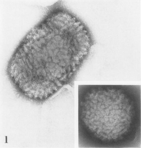
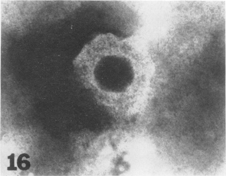
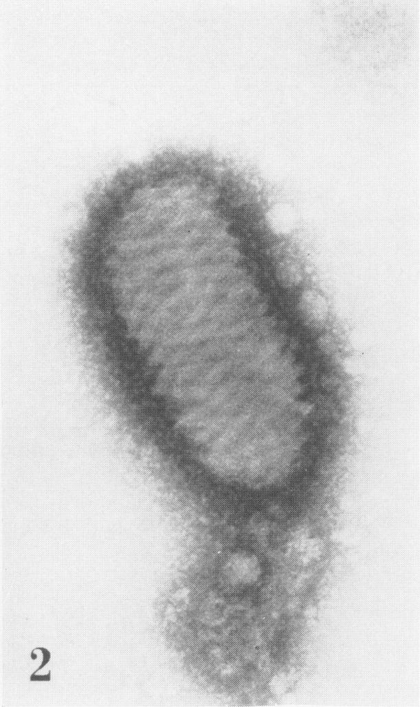
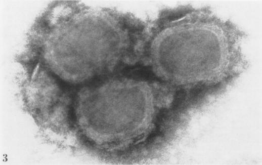
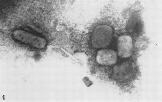
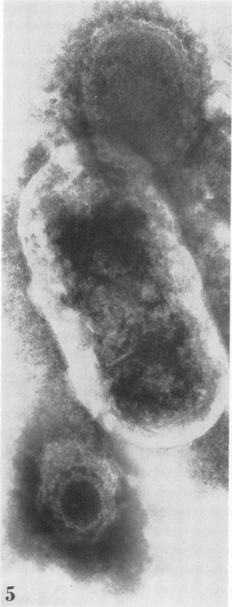
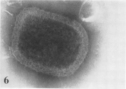
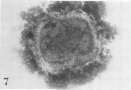
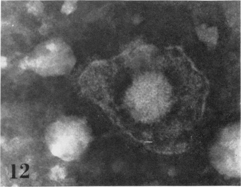
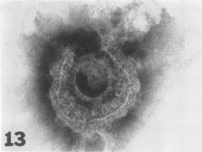
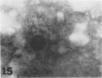
The usefulness of negative-contrast electron microscopy in the rapid differential diagnosis of poxvirus and herpesvirus exanthems is described in this study of 301 specimens from patients with vesicular exanthematous diseases. Specimens from patients with smallpox, various forms of vaccination complications, varicella, zoster (shingles), and herpes simplex are included in this evaluation. Electron microscopy, when applied to the study of lesion material, was found to be more sensitive than the classical techniques of virus isolation in the diagnosis of both poxvirus and herpes/varicella virus infections. However, since specific identification of a virus within a group cannot be made morphologically by electron microscopy, it is recommended that both electron microscopy and virus isolation methods be employed for the routine differential diagnosis of vesicular exanthematous diseases in the reference diagnostic laboratory.
本研究对301例水疱性发疹疾病患者的标本进行了检测,描述了负反差电子显微镜在痘病毒和疱疹病毒疹快速鉴别诊断中的作用。本评估纳入了天花患者、各种形式的疫苗接种并发症患者、水痘患者、带状疱疹(缠腰龙)患者以及单纯疱疹患者的标本。当将电子显微镜应用于病变材料的研究时,发现在痘病毒和疱疹/水痘病毒感染的诊断中,其比传统的病毒分离技术更为敏感。然而,由于无法通过电子显微镜从形态学上对一组病毒进行特异性鉴定,因此建议在参考诊断实验室中,同时采用电子显微镜和病毒分离方法对水疱性发疹疾病进行常规鉴别诊断。