• 文献检索
  • 文档翻译
  • 深度研究
  • 学术资讯
  • Suppr Zotero 插件Zotero 插件
  • 邀请有礼
  • 套餐&价格
  • 历史记录
应用&插件
Suppr Zotero 插件Zotero 插件浏览器插件Mac 客户端Windows 客户端微信小程序
定价
高级版会员购买积分包购买API积分包
服务
文献检索文档翻译深度研究API 文档MCP 服务
关于我们
关于 Suppr公司介绍联系我们用户协议隐私条款
关注我们

Suppr 超能文献

核心技术专利:CN118964589B侵权必究
粤ICP备2023148730 号-1Suppr @ 2026

文献检索

告别复杂PubMed语法,用中文像聊天一样搜索,搜遍4000万医学文献。AI智能推荐,让科研检索更轻松。

立即免费搜索

文件翻译

保留排版,准确专业,支持PDF/Word/PPT等文件格式,支持 12+语言互译。

免费翻译文档

深度研究

AI帮你快速写综述,25分钟生成高质量综述,智能提取关键信息,辅助科研写作。

立即免费体验

艰难梭菌毒素对培养的人羊膜细胞的超微结构变化

Ultrastructural changes of cultured human amnion cells by Clostridiu difficile toxin.

作者信息

Chang T W, Lin P S, Gorbach S L, Bartlett J G

出版信息

Infect Immun. 1979 Mar;23(3):795-8. doi: 10.1128/iai.23.3.795-798.1979.

DOI:10.1128/iai.23.3.795-798.1979
PMID:457259
原文链接:https://pmc.ncbi.nlm.nih.gov/articles/PMC414235/
Abstract

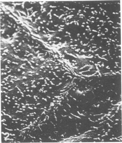
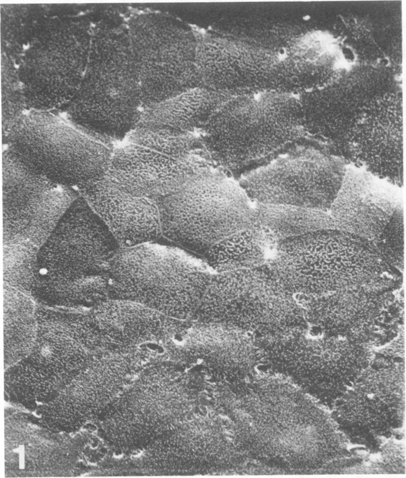
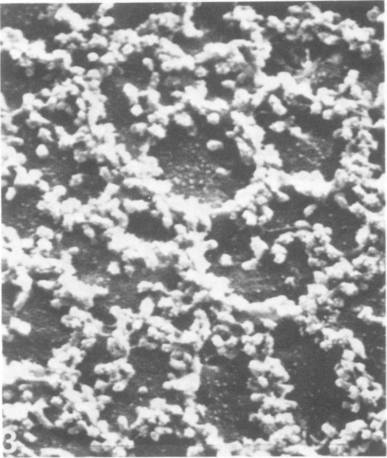
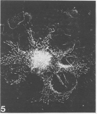
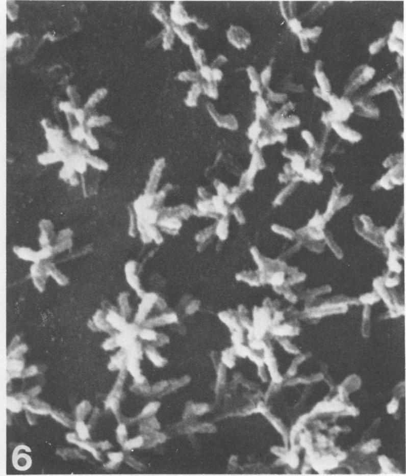
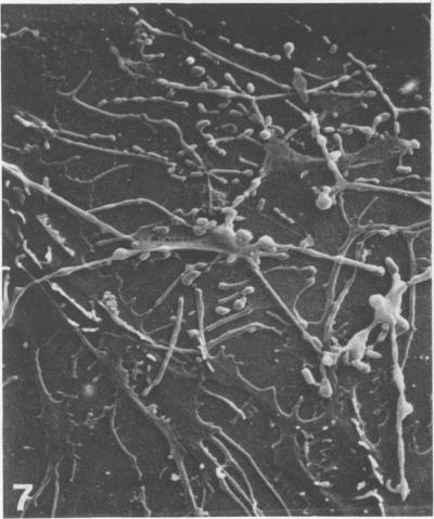
The ultrastructure of the surface of primary human amnion monolayer cells undergoing cytopathology induced by Clostridium difficile toxin was examined by scanning electron microscopy. Our observations indicated that the type and distribution of cell surface projections were altered dramatically by this toxin. The patterns of such surface changes were specific for the two different types of cells found in this cell culture. Cells with demarcated borders showed rearrangement of microvilli into globular chains or ridges which lined up with the branching membrane. Cells without demarcated borders exhibited studlike microvilli, all arranged into ridges or globular chains. These changes were noted after 1 h of toxin exposure and persisted without further progression, in spite of continued toxin exposure, up to 48 h. These data indicate that C. difficile produces a cytolytic toxin and that scanning electron microscopy may be useful in determining toxin-cell interactions.

摘要

利用扫描电子显微镜检查了艰难梭菌毒素诱导细胞病变的原代人羊膜单层细胞表面的超微结构。我们的观察结果表明,这种毒素会显著改变细胞表面突起的类型和分布。这种表面变化模式对于该细胞培养物中发现的两种不同类型的细胞具有特异性。边界清晰的细胞显示微绒毛重排为球状链或脊状,与分支膜排列在一起。边界不清晰的细胞呈现出瘤状微绒毛,全部排列成脊状或球状链。毒素暴露1小时后便观察到这些变化,并且尽管持续暴露于毒素中,这些变化在长达48小时内持续存在且没有进一步发展。这些数据表明艰难梭菌产生一种细胞溶解毒素,并且扫描电子显微镜可能有助于确定毒素与细胞的相互作用。

https://cdn.ncbi.nlm.nih.gov/pmc/blobs/411c/414235/9ba830cbbfe3/iai00183-0245-d.jpg
https://cdn.ncbi.nlm.nih.gov/pmc/blobs/411c/414235/707b4a929de3/iai00183-0244-a.jpg
https://cdn.ncbi.nlm.nih.gov/pmc/blobs/411c/414235/bf52969d3606/iai00183-0244-b.jpg
https://cdn.ncbi.nlm.nih.gov/pmc/blobs/411c/414235/b452ea8e9f4a/iai00183-0244-c.jpg
https://cdn.ncbi.nlm.nih.gov/pmc/blobs/411c/414235/769a4eb8b7cc/iai00183-0244-d.jpg
https://cdn.ncbi.nlm.nih.gov/pmc/blobs/411c/414235/8cf0c313a5e9/iai00183-0245-a.jpg
https://cdn.ncbi.nlm.nih.gov/pmc/blobs/411c/414235/71230604777b/iai00183-0245-b.jpg
https://cdn.ncbi.nlm.nih.gov/pmc/blobs/411c/414235/39412e4ba1df/iai00183-0245-c.jpg
https://cdn.ncbi.nlm.nih.gov/pmc/blobs/411c/414235/9ba830cbbfe3/iai00183-0245-d.jpg

相似文献

1
Ultrastructural changes of cultured human amnion cells by Clostridiu difficile toxin.艰难梭菌毒素对培养的人羊膜细胞的超微结构变化
Infect Immun. 1979 Mar;23(3):795-8. doi: 10.1128/iai.23.3.795-798.1979.
2
Effects of Clostridium difficile toxins A and B in rabbit small and large intestine in vivo and on cultured cells in vitro.艰难梭菌毒素A和B对家兔体内小肠和大肠以及体外培养细胞的作用。
Infect Immun. 1988 Mar;56(3):582-8. doi: 10.1128/iai.56.3.582-588.1988.
3
Clostridium difficile toxin: cytoskeletal changes and lactate dehydrogenase release in hepatocytes.艰难梭菌毒素:肝细胞中的细胞骨架变化及乳酸脱氢酶释放
J Surg Res. 2000 Feb;88(2):165-72. doi: 10.1006/jsre.1999.5736.
4
Saccharomyces boulardii protease inhibits the effects of Clostridium difficile toxins A and B in human colonic mucosa.布拉氏酵母菌蛋白酶可抑制艰难梭菌毒素A和B对人结肠黏膜的作用。
Infect Immun. 1999 Jan;67(1):302-7. doi: 10.1128/IAI.67.1.302-307.1999.
5
EGA Protects Mammalian Cells from Clostridium difficile CDT, Clostridium perfringens Iota Toxin and Clostridium botulinum C2 Toxin.EGA可保护哺乳动物细胞免受艰难梭菌CDT、产气荚膜梭菌iota毒素和肉毒梭菌C2毒素的侵害。
Toxins (Basel). 2016 Apr 1;8(4):101. doi: 10.3390/toxins8040101.
6
Clostridium difficile toxin B is more potent than toxin A in damaging human colonic epithelium in vitro.艰难梭菌毒素B在体外对人结肠上皮细胞的损伤作用比毒素A更强。
J Clin Invest. 1995 May;95(5):2004-11. doi: 10.1172/JCI117885.
7
Clostridium difficile toxins A and B can alter epithelial permeability and promote bacterial paracellular migration through HT-29 enterocytes.艰难梭菌毒素A和B可改变上皮细胞通透性,并促进细菌通过HT-29肠上皮细胞进行旁细胞迁移。
Shock. 2000 Dec;14(6):629-34. doi: 10.1097/00024382-200014060-00010.
8
Effects of Clostridium difficile toxin on tissue-cultured cells.艰难梭菌毒素对组织培养细胞的影响。
J Infect Dis. 1980 Feb;141(2):218-22. doi: 10.1093/infdis/141.2.218.
9
Clostridium difficile Toxin Biology.艰难梭菌毒素生物学。
Annu Rev Microbiol. 2017 Sep 8;71:281-307. doi: 10.1146/annurev-micro-090816-093458. Epub 2017 Jun 28.
10
Ultrastructural effects of Clostridium difficile toxin B on smooth muscle cells and fibroblasts.艰难梭菌毒素B对平滑肌细胞和成纤维细胞的超微结构影响
Exp Cell Res. 1983 Oct 15;148(2):413-22. doi: 10.1016/0014-4827(83)90163-5.

引用本文的文献

1
infection: traversing host-pathogen interactions in the gut.感染:穿越肠道中的宿主-病原体相互作用。
Microbiology (Reading). 2023 Feb;169(2). doi: 10.1099/mic.0.001306.
2
Recent Advances in the Diagnosis and Treatment of Clostridium Difficile Infection.艰难梭菌感染的诊断与治疗新进展
F1000Res. 2016 Jan 29;5. doi: 10.12688/f1000research.7109.1. eCollection 2016.
3
Identification of Clostridium difficile toxin B cardiotoxicity using a zebrafish embryo model of intoxication.利用斑马鱼胚胎中毒模型鉴定艰难梭菌毒素B的心脏毒性

本文引用的文献

1
Binding of human lymphotoxin to target-cell membranes and its relation to cell-mediated cytodestruction.
Proc Natl Acad Sci U S A. 1973 Nov;70(11):3082-6. doi: 10.1073/pnas.70.11.3082.
2
The effects of ophthalmic drugs, vehicles, and preservatives on corneal epithelium: a scanning electron microscope study.眼科药物、赋形剂和防腐剂对角膜上皮的影响:一项扫描电子显微镜研究。
Invest Ophthalmol. 1976 Apr;15(4):246-59.
3
Comparison of cancer cell surfaces of the lower reproductive tract by scanning electron microscopy.
Am J Obstet Gynecol. 1977 Dec 15;129(8):893-908. doi: 10.1016/0002-9378(77)90522-1.
4
Proc Natl Acad Sci U S A. 2006 Sep 19;103(38):14176-81. doi: 10.1073/pnas.0604725103. Epub 2006 Sep 11.
4
Clostridium difficile toxins: mechanism of action and role in disease.艰难梭菌毒素:作用机制及在疾病中的作用
Clin Microbiol Rev. 2005 Apr;18(2):247-63. doi: 10.1128/CMR.18.2.247-263.2005.
5
Neutralizing activity against Clostridium difficile toxin in the supernatants of cultured colostral cells.培养的初乳细胞上清液中针对艰难梭菌毒素的中和活性。
Infect Immun. 1980 Aug;29(2):545-50. doi: 10.1128/iai.29.2.545-550.1980.
6
Mucosal damage mediated by clostridial toxin in experimental clindamycin-associated colitis.实验性克林霉素相关性结肠炎中梭菌毒素介导的黏膜损伤
Gut. 1980 Jun;21(6):493-9. doi: 10.1136/gut.21.6.493.
7
Loss of surface fibronectin from human lung fibroblasts exposed to cytotoxin from Clostridium difficile.暴露于艰难梭菌细胞毒素的人肺成纤维细胞表面纤连蛋白的丧失
Infect Immun. 1983 Mar;39(3):1470-2. doi: 10.1128/iai.39.3.1470-1472.1983.
8
Cell surface binding site for Clostridium difficile enterotoxin: evidence for a glycoconjugate containing the sequence Gal alpha 1-3Gal beta 1-4GlcNAc.艰难梭菌肠毒素的细胞表面结合位点:含序列Galα1-3Galβ1-4GlcNAc的糖缀合物的证据
Infect Immun. 1986 Sep;53(3):573-81. doi: 10.1128/iai.53.3.573-581.1986.
9
Laboratory diagnosis of Clostridium difficile-associated diarrhoea.艰难梭菌相关性腹泻的实验室诊断
Eur J Clin Microbiol Infect Dis. 1988 Aug;7(4):476-84. doi: 10.1007/BF01962596.
Clindamycin-associated colitis due to a toxin-producing species of Clostridium in hamsters.仓鼠中由产毒素梭状芽孢杆菌引起的克林霉素相关性结肠炎。
J Infect Dis. 1977 Nov;136(5):701-5. doi: 10.1093/infdis/136.5.701.
5
Clindamycin-induced enterocolitis in hamsters as a model of pseudomembranous colitis in patients.以仓鼠克林霉素诱导的小肠结肠炎作为患者伪膜性结肠炎的模型。
Infect Immun. 1978 May;20(2):526-9. doi: 10.1128/iai.20.2.526-529.1978.
6
Identification of Clostridium difficile as a cause of pseudomembranous colitis.艰难梭菌被鉴定为伪膜性结肠炎的病因。
Br Med J. 1978 Mar 18;1(6114):695. doi: 10.1136/bmj.1.6114.695.
7
Antibiotic-associated pseudomembranous colitis due to toxin-producing clostridia.由产毒素梭状芽孢杆菌引起的抗生素相关性假膜性结肠炎。
N Engl J Med. 1978 Mar 9;298(10):531-4. doi: 10.1056/NEJM197803092981003.
8
Clostridium difficile and the aetiology of pseudomembranous colitis.艰难梭菌与伪膜性结肠炎的病因学
Lancet. 1978 May 20;1(8073):1063-6. doi: 10.1016/s0140-6736(78)90912-1.
9
Antibiotic-induced colitis implication of a toxin neutralised by Clostridium sordellii antitoxin.抗生素诱导的结肠炎与一种被索氏梭菌抗毒素中和的毒素有关。
Lancet. 1977 Nov 26;2(8048):1103-6. doi: 10.1016/s0140-6736(77)90547-5.
10
Cyclic amp and cell morphology in cultured fibroblasts. Effects on cell shape, microfilament and microtubule distribution, and orientation to substratum.培养的成纤维细胞中的环磷酸腺苷与细胞形态。对细胞形状、微丝和微管分布以及与基质的取向的影响。
J Cell Biol. 1975 Oct;67(1):146-59. doi: 10.1083/jcb.67.1.146.