• 文献检索
  • 文档翻译
  • 深度研究
  • 学术资讯
  • Suppr Zotero 插件Zotero 插件
  • 邀请有礼
  • 套餐&价格
  • 历史记录
应用&插件
Suppr Zotero 插件Zotero 插件浏览器插件Mac 客户端Windows 客户端微信小程序
定价
高级版会员购买积分包购买API积分包
服务
文献检索文档翻译深度研究API 文档MCP 服务
关于我们
关于 Suppr公司介绍联系我们用户协议隐私条款
关注我们

Suppr 超能文献

核心技术专利:CN118964589B侵权必究
粤ICP备2023148730 号-1Suppr @ 2026

文献检索

告别复杂PubMed语法,用中文像聊天一样搜索,搜遍4000万医学文献。AI智能推荐,让科研检索更轻松。

立即免费搜索

文件翻译

保留排版,准确专业,支持PDF/Word/PPT等文件格式,支持 12+语言互译。

免费翻译文档

深度研究

AI帮你快速写综述,25分钟生成高质量综述,智能提取关键信息,辅助科研写作。

立即免费体验

鼻硬结病中的米库利奇细胞。光镜、荧光镜及电镜研究。

The Mikulicz cell in rhinoscleroma. Light, fluorescent and electron microscopic studies.

作者信息

Hoffmann E O, Loose L D, Harkin J C

出版信息

Am J Pathol. 1973 Oct;73(1):47-58.

PMID:4584128
原文链接:https://pmc.ncbi.nlm.nih.gov/articles/PMC1904057/
Abstract

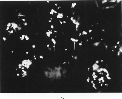
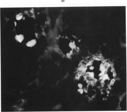
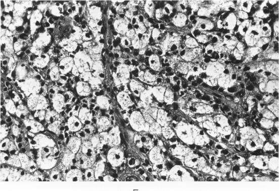
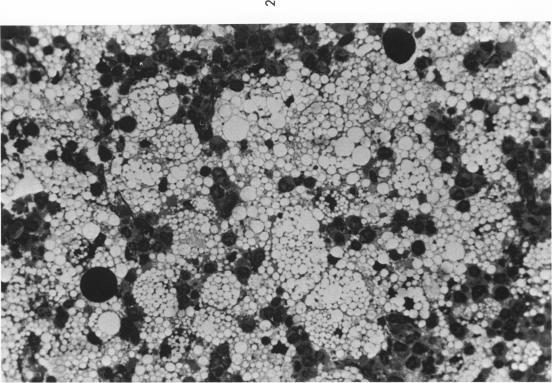
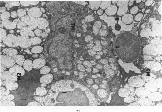
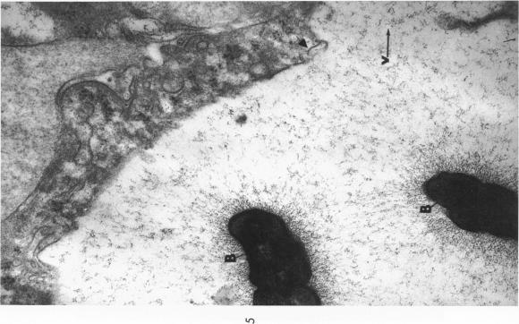
The stages in the development of the Mikulicz cell in human rhinoscleroma were studied in biopsy specimens obtained from 10 patients using light, immunofluorescent and electron microscopy. The Mikulicz cell was identified morphologically as a macrophage, not a plasma cell. Acutely inflamed areas of rhinoscleroma presented abundant bacteria with a slime layer. The microorganism was infrequent and the mucopolysaccharide was scanty in rhinoscleromal tissue, where plasma cells predominated, and in cicatricial fibrous tissue. In the granulomatous stage of rhinoscleroma, the mucopolysaccharide was found within the Mikulicz cells. The vacuoles observed in the Mikulicz cells were considered to be phagosomes containing, principally, bacterial mucopolysaccharide and few bacteria and, to a lesser extent, swollen mitochondria. It was concluded that the slime layer of Klebsiella rhinoscleromatis plays an important role in the pathogenesis of the disease. It is postulated that this material is a nondigestible mucopolysaccharide that resides in the phagosomes of macrophages, increases the osmotic pressure and forms multiple hydropic vacuoles that rupture not only the phagosomes but also the cells, resulting in the liberation of the mucopolysaccharide. This would initiate a cycle that would prolong the disease in the absence of the bacteria.

摘要

利用光学显微镜、免疫荧光显微镜和电子显微镜,对10例人鼻硬结病患者活检标本中米库利奇细胞(Mikulicz cell)的发育阶段进行了研究。从形态学上看,米库利奇细胞被确定为巨噬细胞,而非浆细胞。鼻硬结病的急性炎症区域可见大量带有黏液层的细菌。在以浆细胞为主的鼻硬结病组织及瘢痕纤维组织中,微生物较少,黏多糖也很少。在鼻硬结病的肉芽肿阶段,在米库利奇细胞内发现了黏多糖。米库利奇细胞中观察到的空泡被认为是吞噬体,主要含有细菌黏多糖和少量细菌,其次还含有肿胀的线粒体。得出的结论是,鼻硬结克雷伯菌的黏液层在该病的发病机制中起重要作用。据推测,这种物质是一种不可消化的黏多糖,存在于巨噬细胞的吞噬体内,增加渗透压并形成多个水泡样空泡,这些空泡不仅会使吞噬体破裂,还会使细胞破裂,导致黏多糖释放。在没有细菌的情况下,这将引发一个使疾病持续的循环。

https://cdn.ncbi.nlm.nih.gov/pmc/blobs/ee3e/1904057/0d1f95fb6c86/amjpathol00248-0061-b.jpg
https://cdn.ncbi.nlm.nih.gov/pmc/blobs/ee3e/1904057/160b4c17f9c7/amjpathol00248-0062-a.jpg
https://cdn.ncbi.nlm.nih.gov/pmc/blobs/ee3e/1904057/df4e5852bfb8/amjpathol00248-0062-b.jpg
https://cdn.ncbi.nlm.nih.gov/pmc/blobs/ee3e/1904057/648c04645006/amjpathol00248-0059-a.jpg
https://cdn.ncbi.nlm.nih.gov/pmc/blobs/ee3e/1904057/e403bdea4920/amjpathol00248-0059-b.jpg
https://cdn.ncbi.nlm.nih.gov/pmc/blobs/ee3e/1904057/99b1b3e323bd/amjpathol00248-0060-a.jpg
https://cdn.ncbi.nlm.nih.gov/pmc/blobs/ee3e/1904057/a8e05fa913ed/amjpathol00248-0060-b.jpg
https://cdn.ncbi.nlm.nih.gov/pmc/blobs/ee3e/1904057/a9d6fcfa8865/amjpathol00248-0061-a.jpg
https://cdn.ncbi.nlm.nih.gov/pmc/blobs/ee3e/1904057/0d1f95fb6c86/amjpathol00248-0061-b.jpg

相似文献

1
The Mikulicz cell in rhinoscleroma. Light, fluorescent and electron microscopic studies.鼻硬结病中的米库利奇细胞。光镜、荧光镜及电镜研究。
Am J Pathol. 1973 Oct;73(1):47-58.
2
[Electron microscopical investigation of rhinoscleroma (author's transl)].鼻硬结病的电子显微镜研究(作者译)
Arch Dermatol Res (1975). 1975 Dec 31;254(3):263-74. doi: 10.1007/BF00557968.
3
[Light and electron microscopy of rhinoscleroma (author's transl)].鼻硬结病的光镜和电镜检查(作者译)
HNO. 1975 Feb;23(2):35-42.
4
The role of plasma cells in scleroma. Electron-microscopic study.浆细胞在硬结症中的作用。电子显微镜研究。
ORL J Otorhinolaryngol Relat Spec. 1977;39(1):1-13. doi: 10.1159/000275333.
5
Cytologic diagnosis of rhinoscleroma.鼻硬结病的细胞学诊断
Acta Cytol. 1984 Mar-Apr;28(2):139-42.
6
[An etiological and pathologic study of Rhinoscleroma].[鼻硬结病的病因及病理学研究]
Zhonghua Bing Li Xue Za Zhi. 2000 Dec;29(6):421-3.
7
[Clinicopathologic analysis of rhinoscleroma].[鼻硬结病的临床病理分析]
Zhonghua Er Bi Yan Hou Ke Za Zhi. 2001 Feb;36(1):42-3.
8
Rhinoscleroma pathogenesis: The type K3 capsule of Klebsiella rhinoscleromatis is a virulence factor not involved in Mikulicz cells formation.类鼻疽发病机制:类鼻疽伯克霍尔德菌 K 型荚膜是一种毒力因子,不参与米库利兹细胞的形成。
PLoS Negl Trop Dis. 2018 Jan 30;12(1):e0006201. doi: 10.1371/journal.pntd.0006201. eCollection 2018 Jan.
9
Nasal rhinoscleroma in a nonendemic area: a case report.非流行地区的鼻硬结病:一例报告
J Korean Med Sci. 2003 Jun;18(3):455-8. doi: 10.3346/jkms.2003.18.3.455.
10
An interpretation of the structural changes responsible for the chronicity of rhinoscleroma.对导致鼻硬结病慢性化的结构变化的一种解读。
Laryngoscope. 2001 Jun;111(6):1020-6. doi: 10.1097/00005537-200106000-00016.

引用本文的文献

1
Rhinoscleroma: a detailed histopathological diagnostic insight.鼻硬结病:详细的组织病理学诊断见解
Int J Clin Exp Pathol. 2015 Jul 1;8(7):8438-45. eCollection 2015.
2
Rhinoscleroma: a case series report and review of the literature.鼻硬结病:病例系列报告及文献综述
Eur Arch Otorhinolaryngol. 2014 Jul;271(7):1851-6. doi: 10.1007/s00405-013-2649-z. Epub 2013 Aug 1.
3
A novel murine model of rhinoscleroma identifies Mikulicz cells, the disease signature, as IL-10 dependent derivatives of inflammatory monocytes.一种新型的鼻硬结病鼠模型将 Mikulicz 细胞(疾病标志物)鉴定为依赖于 IL-10 的炎症单核细胞衍生细胞。

本文引用的文献

1
IN VIVO AND IN VITRO OBSERVATIONS OF LEPTOSPIRA POMONA BY ELECTRON MICROSCOPY.通过电子显微镜对波摩那钩端螺旋体进行的体内和体外观察
J Bacteriol. 1962 Sep;84(3):569-76. doi: 10.1128/jb.84.3.569-576.1962.
2
OCCURRENCE OF PYRUVIC ACID IN THE CAPSULAR POLYSACCHARIDE OF KLEBSIELLA RHINOSCLEROMATIS.鼻硬结克雷伯菌荚膜多糖中丙酮酸的存在情况
J Bacteriol. 1965 Feb;89(2):539. doi: 10.1128/jb.89.2.539-539.1965.
3
RHINOSCLEROMA. LIGHT AND ELECTRON MICROSCOPIC STUDIES.鼻硬结病。光镜和电镜研究。
EMBO Mol Med. 2013 Apr;5(4):516-30. doi: 10.1002/emmm.201202023.
4
Infectious pseudotumors: red herrings in head and neck pathology.感染性假瘤:头颈部病理学中的干扰因素
Head Neck Pathol. 2012 Mar;6(1):58-63. doi: 10.1007/s12105-012-0340-0. Epub 2012 Mar 20.
5
Septal rhinoscleroma.鼻中隔硬结病
Indian J Plast Surg. 2010 Jul;43(2):219-21. doi: 10.4103/0970-0358.73465.
6
A case of rhinoscleroma cured with ciprofloxacin.1例用环丙沙星治愈的鼻硬结病。
Infection. 1993 Nov-Dec;21(6):403-6. doi: 10.1007/BF01728925.
7
Alterations in the T-lymphocyte subpopulation in patients with rhinoscleroma.鼻硬结病患者T淋巴细胞亚群的改变。
J Clin Microbiol. 1988 May;26(5):1031-3. doi: 10.1128/jcm.26.5.1031-1033.1988.
8
Scleroma of the nose and pharynx.鼻与咽硬结病
West J Med. 1976 Jan;124(1):13-7.
9
[Electron microscopical investigation of rhinoscleroma (author's transl)].鼻硬结病的电子显微镜研究(作者译)
Arch Dermatol Res (1975). 1975 Dec 31;254(3):263-74. doi: 10.1007/BF00557968.
Arch Pathol. 1964 Nov;78:501-12.
4
ELECTRON MICROSCOPE OBSERVATIONS OF BRUCELLA ABORTUS GROWN WITHIN MONOCYTES IN VITRO.布鲁氏菌流产亚种在体外单核细胞内生长的电子显微镜观察
J Gen Microbiol. 1964 Jun;35:383-90. doi: 10.1099/00221287-35-3-383.
5
Light and electron microscopic observations of scleroma.硬结症的光学显微镜和电子显微镜观察
Exp Mol Pathol. 1963 Feb;2:93-101. doi: 10.1016/0014-4800(63)90011-x.
6
Rhinoscleroma--a clinical perspective.鼻硬结病——临床视角
J Laryngol Otol. 1961 Dec;75:1011-39. doi: 10.1017/s0022215100058837.
7
Electron microscopic study of Histoplasma in mouse spleen.小鼠脾脏中荚膜组织胞浆菌的电子显微镜研究。
J Bacteriol. 1959 Apr;77(4):429-38. doi: 10.1128/jb.77.4.429-438.1959.
8
Scleroma respiratorium in Indonesia, particularly the scleroma endemic in North Celebes.印度尼西亚的呼吸道硬结病,尤其是北苏拉威西岛特有的地方性硬结病。
Doc Med Geogr Trop. 1956 Jun;8(2):101-16.
9
Electron microscopic studies of scleroma granulation tissue.硬结症肉芽组织的电子显微镜研究。
Acta Med Pol. 1969;10(2):231-42.
10
The uptake, storage, and intracellular hydrolysis of carbohydrates by macrophages.巨噬细胞对碳水化合物的摄取、储存及细胞内水解作用。
J Exp Med. 1969 Jan 1;129(1):201-25. doi: 10.1084/jem.129.1.201.