• 文献检索
  • 文档翻译
  • 深度研究
  • 学术资讯
  • Suppr Zotero 插件Zotero 插件
  • 邀请有礼
  • 套餐&价格
  • 历史记录
应用&插件
Suppr Zotero 插件Zotero 插件浏览器插件Mac 客户端Windows 客户端微信小程序
定价
高级版会员购买积分包购买API积分包
服务
文献检索文档翻译深度研究API 文档MCP 服务
关于我们
关于 Suppr公司介绍联系我们用户协议隐私条款
关注我们

Suppr 超能文献

核心技术专利:CN118964589B侵权必究
粤ICP备2023148730 号-1Suppr @ 2026

文献检索

告别复杂PubMed语法,用中文像聊天一样搜索,搜遍4000万医学文献。AI智能推荐,让科研检索更轻松。

立即免费搜索

文件翻译

保留排版,准确专业,支持PDF/Word/PPT等文件格式,支持 12+语言互译。

免费翻译文档

深度研究

AI帮你快速写综述,25分钟生成高质量综述,智能提取关键信息,辅助科研写作。

立即免费体验

正常淋巴细胞亚群的精细结构——一项使用单克隆抗体和免疫金技术的研究

The fine structure of normal lymphocyte subpopulations--a study with monoclonal antibodies and the immunogold technique.

作者信息

Matutes E, Catovsky D

出版信息

Clin Exp Immunol. 1982 Nov;50(2):416-25.

PMID:6185261
原文链接:https://pmc.ncbi.nlm.nih.gov/articles/PMC1536696/
Abstract

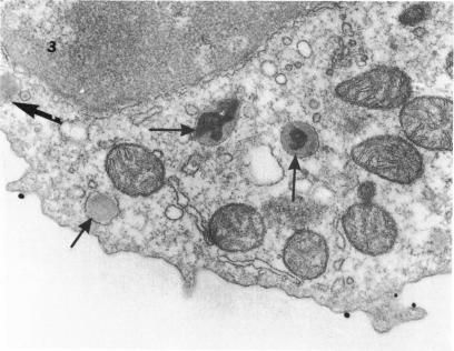
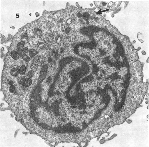
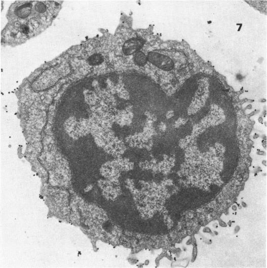
The ultrastructural characteristics of normal lymphocyte subpopulations, identified by monoclonal antibodies and visualized by a colloidal gold labelled anti-mouse IgG were analysed. Our study demonstrates: (1) the major T lymphocyte subsets (OKT4+ and OKT8+) have distinct ultrastructural morphology. The majority of OKT4+ cells have a high nuclear/cytoplasmic ratio (N/C) and few cytoplasmic organelles whilst most OKT8+ cells have a low N/C ratio and numerous organelles, namely a well developed Golgi apparatus, lysosomal granules and parallel tubular arrays (PTA); (2) a unique subtype with irregular nuclear outline that resembles Sézary cells was seen in 5-10% of OKT4+ lymphocytes; (3) OKM1, a reagent that reacts with monocytes and granulocytes, is positive in a small lymphocyte subset which appears to be negative with the OKT reagents and is morphologically identical to OKT8+ cells; (4) 'hand-mirror' cells were only seen labelled with OKT8 and OKM1; (5) B lymphocytes labelled with FMC4 (anti-IA) could be distinguished from OKT3+ lymphocytes by having numerous profiles of endoplasmic reticulum (ER) and ribosomes; these were particularly prominent in lymphoplasmacytoid cells. Morphological similarities between normal T lymphocyte subsets and T neoplasias of the same membrane phenotype suggest that these disorders arise from specific T cell types present in normal peripheral blood or from common precursors.

摘要

分析了通过单克隆抗体鉴定并用胶体金标记的抗小鼠IgG可视化的正常淋巴细胞亚群的超微结构特征。我们的研究表明:(1)主要的T淋巴细胞亚群(OKT4 +和OKT8 +)具有不同的超微结构形态。大多数OKT4 +细胞具有高核/质比(N/C)且细胞质细胞器较少,而大多数OKT8 +细胞具有低N/C比且细胞器众多,即发达的高尔基体、溶酶体颗粒和平行管状排列(PTA);(2)在5%-10%的OKT4 +淋巴细胞中可见一种具有不规则核轮廓且类似于Sezary细胞的独特亚型;(3)OKM1是一种与单核细胞和粒细胞反应的试剂,在一个小淋巴细胞亚群中呈阳性,该亚群似乎对OKT试剂呈阴性,且在形态上与OKT8 +细胞相同;(4)“手镜”细胞仅用OKT8和OKM1标记;(5)用FMC4(抗-IA)标记的B淋巴细胞可通过具有大量内质网(ER)和核糖体轮廓与OKT3 +淋巴细胞区分开来;这些在淋巴浆细胞样细胞中尤为突出。正常T淋巴细胞亚群与相同膜表型的T肿瘤之间的形态学相似性表明,这些疾病源于正常外周血中存在的特定T细胞类型或共同前体。

https://cdn.ncbi.nlm.nih.gov/pmc/blobs/d250/1536696/7d765586293e/clinexpimmunol00164-0186-b.jpg
https://cdn.ncbi.nlm.nih.gov/pmc/blobs/d250/1536696/fe932178ec3c/clinexpimmunol00164-0182-a.jpg
https://cdn.ncbi.nlm.nih.gov/pmc/blobs/d250/1536696/fde3343b3969/clinexpimmunol00164-0183-a.jpg
https://cdn.ncbi.nlm.nih.gov/pmc/blobs/d250/1536696/42ed807ed06e/clinexpimmunol00164-0183-b.jpg
https://cdn.ncbi.nlm.nih.gov/pmc/blobs/d250/1536696/2971796a351e/clinexpimmunol00164-0184-a.jpg
https://cdn.ncbi.nlm.nih.gov/pmc/blobs/d250/1536696/cda047e1f1d6/clinexpimmunol00164-0184-b.jpg
https://cdn.ncbi.nlm.nih.gov/pmc/blobs/d250/1536696/b3741216bf0c/clinexpimmunol00164-0185-a.jpg
https://cdn.ncbi.nlm.nih.gov/pmc/blobs/d250/1536696/b8d01476f13b/clinexpimmunol00164-0185-b.jpg
https://cdn.ncbi.nlm.nih.gov/pmc/blobs/d250/1536696/f642a63028ab/clinexpimmunol00164-0186-a.jpg
https://cdn.ncbi.nlm.nih.gov/pmc/blobs/d250/1536696/7d765586293e/clinexpimmunol00164-0186-b.jpg

相似文献

1
The fine structure of normal lymphocyte subpopulations--a study with monoclonal antibodies and the immunogold technique.正常淋巴细胞亚群的精细结构——一项使用单克隆抗体和免疫金技术的研究
Clin Exp Immunol. 1982 Nov;50(2):416-25.
2
T lymphocytic subpopulations of normal human peripheral blood defined by monoclonal antibodies: an ultrastructural study using immunogold staining method.单克隆抗体界定的正常人外周血T淋巴细胞亚群:应用免疫金染色法的超微结构研究
Basic Appl Histochem. 1986;30(1):41-52.
3
Human thymocyte differentiation: an ultrastructural study using monoclonal antibodies and immunogold staining method.人类胸腺细胞分化:一项使用单克隆抗体和免疫金染色法的超微结构研究。
Biomed Pharmacother. 1986;40(7):253-60.
4
Characterization of the alloactivated E+Ia+ cell capable of stimulating in mixed lymphocyte culture.能够在混合淋巴细胞培养中发挥刺激作用的同种异体活化E+Ia+细胞的特性分析。
J Immunol. 1982 Jan;128(1):501-5.
5
Analysis of Fc (IgG) receptors on human peripheral blood leukocytes by dual fluorescence flow microfluorometry. II. Quantitation of receptors on cells that express the OKM1, OKT3, OKT4, and OKT8 antigens.采用双荧光流式微量荧光测定法分析人外周血白细胞上的Fc(IgG)受体。II. 表达OKM1、OKT3、OKT4和OKT8抗原的细胞上受体的定量分析。
J Immunol. 1983 Mar;130(3):1152-8.
6
Morphological heterogeneity of Leu7, Leu11 and OKM1 positive lymphocyte subsets: an ultrastructural study with the immunogold method.Leu7、Leu11和OKM1阳性淋巴细胞亚群的形态学异质性:免疫金法的超微结构研究
Clin Exp Immunol. 1987 May;68(2):331-9.
7
Candidate counterparts of Sézary cells and adult T-cell lymphoma-leukaemia cells in normal peripheral blood: an ultrastructural study with the immunogold method and monoclonal antibodies.正常外周血中Sezary细胞和成人T细胞淋巴瘤白血病细胞的候选对应物:免疫金法和单克隆抗体的超微结构研究
Leuk Res. 1983;7(6):787-801. doi: 10.1016/0145-2126(83)90073-5.
8
Immunoelectron microscopic application of monoclonal antibodies for identification of lymphocyte subsets bearing tubuloreticular inclusions or parallel tubular arrays.单克隆抗体在免疫电子显微镜下的应用,用于鉴定带有管状网状包涵体或平行管状排列的淋巴细胞亚群。
J Exp Pathol. 1984;1(2):157-73.
9
Synovial fluid lymphocytes differ from peripheral blood lymphocytes in patients with rheumatoid arthritis.类风湿性关节炎患者的滑液淋巴细胞与外周血淋巴细胞不同。
J Immunol. 1982 Jan;128(1):351-4.
10
Differential recovery of circulating T cell subsets after nodal irradiation for Hodgkin's disease.霍奇金病淋巴结照射后循环T细胞亚群的差异恢复
J Immunol. 1984 Feb;132(2):1026-30.

引用本文的文献

1
Identification of Fas-L-expressing apoptotic T lymphocytes in normal human peripheral blood : in vivo suicide.正常人外周血中表达Fas-L的凋亡性T淋巴细胞的鉴定:体内自杀
Am J Pathol. 2001 Feb;158(2):387-91. doi: 10.1016/S0002-9440(10)63981-8.
2
Ultrastructural cytochemistry of chronic T-cell leukaemias. A study with four acid hydrolases.
Histochem J. 1983 Sep;15(9):895-909. doi: 10.1007/BF01011828.
3
The fine structure of HNK-1 (Leu7) positive cells. A study using an immunoperoxidase technique.HNK-1(Leu7)阳性细胞的精细结构。一项采用免疫过氧化物酶技术的研究。

本文引用的文献

1
Unusual phenotype and function of an expanded subpopulation of T cells in patients with haemopoietic disorders.造血系统疾病患者中T细胞亚群扩增的异常表型与功能
Clin Exp Immunol. 1981 Mar;43(3):497-505.
2
A monoclonal antibody reactive with human peripheral blood monocytes.一种与人外周血单核细胞反应的单克隆抗体。
J Immunol. 1980 Apr;124(4):1943-8.
3
Morphological aspects of T cell subpopulations in human blood: characterization of the cerebriform mononuclear cells in healthy individuals.人血中T细胞亚群的形态学特征:健康个体中脑形单核细胞的特性
Histochemistry. 1984;81(2):153-5. doi: 10.1007/BF00490109.
4
The use of a peroxidase-anti-peroxidase complex for the visualization of monoclonal antibodies on the ultrastructural level.使用过氧化物酶-抗过氧化物酶复合物在超微结构水平上显示单克隆抗体。
Clin Exp Immunol. 1983 Dec;54(3):819-26.
5
A novel approach for scanning electron microscopy of colloidal gold-labeled cell surfaces.一种用于胶体金标记细胞表面扫描电子显微镜观察的新方法。
J Cell Biol. 1984 Jul;99(1 Pt 1):53-7. doi: 10.1083/jcb.99.1.53.
6
Leu-7-positive cells with monocyte phenotype show different ultrastructural features in comparison to Leu-7-positive cells with T-cell phenotype.与具有T细胞表型的Leu-7阳性细胞相比,具有单核细胞表型的Leu-7阳性细胞表现出不同的超微结构特征。
Histochemistry. 1985;82(6):525-7. doi: 10.1007/BF00489973.
7
Morphological heterogeneity of Leu7, Leu11 and OKM1 positive lymphocyte subsets: an ultrastructural study with the immunogold method.Leu7、Leu11和OKM1阳性淋巴细胞亚群的形态学异质性:免疫金法的超微结构研究
Clin Exp Immunol. 1987 May;68(2):331-9.
8
An unusual variant of T-CLL: evidence for the existence of a hitherto unrecognized T cell subset.一种不寻常的T细胞慢性淋巴细胞白血病变体:存在迄今未被识别的T细胞亚群的证据。
Clin Exp Immunol. 1986 Feb;63(2):303-11.
9
Regulatory peptide immunocytochemistry at light- and electron microscopical levels.光镜和电镜水平下的调节肽免疫细胞化学
Experientia. 1987 Jul 15;43(7):724-34. doi: 10.1007/BF01945349.
10
Correlated ultrastructural and immunological findings in CD8+ and CD4+ peripheral T-cell lymphomas.CD8 +和CD4 +外周T细胞淋巴瘤的相关超微结构和免疫学发现
Med Oncol Tumor Pharmacother. 1988;5(3):173-80. doi: 10.1007/BF02986441.
Clin Exp Immunol. 1981 Mar;43(3):506-16.
4
Chronic T-cell leukemias. I. Morphology, cytochemistry and ultrastructure.
Leuk Res. 1980;4(5):463-76. doi: 10.1016/0145-2126(80)90028-4.
5
Phenotypic characterization of cutaneous T-cell lymphoma. Use of monoclonal antibodies to compare with other malignant T cells.皮肤T细胞淋巴瘤的表型特征。使用单克隆抗体与其他恶性T细胞进行比较。
N Engl J Med. 1981 May 28;304(22):1319-23. doi: 10.1056/NEJM198105283042202.
6
Ultrastructural localization of acid phosphatase in rosetted T and B lymphocytes of normal human blood.正常人血中玫瑰花结形成的T淋巴细胞和B淋巴细胞酸性磷酸酶的超微结构定位
Am J Pathol. 1981 Jan;102(1):72-83.
7
Human T lymphocytes of inducer and suppressor type occupy different microenvironments.诱导型和抑制型人类T淋巴细胞占据不同的微环境。
Nature. 1980 Nov 6;288(5786):81-4. doi: 10.1038/288081a0.
8
Evidence by reactivity with hybridoma antibodies for a probable myeloid origin of peripheral blood cells active in natural cytotoxicity and antibody-dependent cell-mediated cytotoxicity.与杂交瘤抗体反应的证据表明,参与自然细胞毒性和抗体依赖性细胞介导的细胞毒性的外周血细胞可能起源于髓系。
J Clin Invest. 1980 Oct;66(4):847-51. doi: 10.1172/JCI109923.
9
Human lymphocyte markers defined by antibodies derived from somatic cell hybrids. II. A hybridoma secreting antibody against an antigen expressed by human B and null lymphocytes.由体细胞杂交产生的抗体所定义的人类淋巴细胞标志物。II. 一种分泌针对人类B淋巴细胞和裸淋巴细胞所表达抗原的抗体的杂交瘤。
Clin Exp Immunol. 1980 Jun;40(3):593-601.
10
Ultrastructure and cytochemistry of human peripheral blood lymphocytes. Similarities between the cells of the third population and TG lymphocytes.人外周血淋巴细胞的超微结构与细胞化学。第三群细胞与TG淋巴细胞之间的相似性。
Eur J Immunol. 1980 Jul;10(7):562-70. doi: 10.1002/eji.1830100714.