Kirkpatrick J N, Manivannan A, Gupta A K, Hipwell J, Forrester J V, Sharp P F
University of Aberdeen, Medical School, Foresterhill.
Br J Ophthalmol. 1995 Oct;79(10):892-9. doi: 10.1136/bjo.79.10.892.
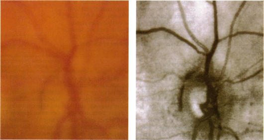
An investigation was carried out to compare the image quality of the ocular fundus obtained clinically, photographically, and with the scanning laser ophthalmoscope (SLO) at visible and infrared wavelengths in patients with significant cataract.
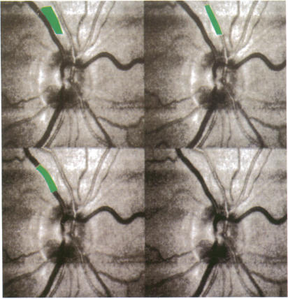
Nineteen patients admitted for routine cataract extraction were examined clinically by two independent observers to ascertain cataract type and clarity of fundus view with an indirect ophthalmoscope. Fundus photography and both confocal and direct (non-confocal) SLO imaging at 590 nm, 670 nm, and 830 nm were carried out after pupillary dilatation. Images obtained were graded independently using a recognised grading system.
Quality of SLO images appeared to be superior to indirect ophthalmoscopy (p < 0.01) and fundus photography (p < 0.001) when graded subjectively. Quantitative analysis of contrast of retinal vessels demonstrated significantly higher contrast for the SLO compared with digitised fundus photographs at all wavelengths tested (p < 0.001), with highest contrast at 590 nm. Use of a confocal aperture significantly improved vessel contrast but may reduce overall image intensity.
Scanning laser ophthalmoscopy may offer a method to observe and record fine fundus detail in patients who have marked cataract.
开展一项研究,比较在患有明显白内障的患者中,通过临床检查、眼底照相以及使用扫描激光检眼镜(SLO)在可见光和红外波长下所获得的眼底图像质量。
19例因常规白内障摘除术入院的患者由两名独立观察者进行临床检查,以通过间接检眼镜确定白内障类型和眼底观察的清晰度。在瞳孔散大后进行眼底照相以及在590nm、670nm和830nm波长下的共焦和直接(非共焦)SLO成像。所获得的图像使用公认的分级系统进行独立分级。
主观分级时,SLO图像质量似乎优于间接检眼镜检查(p < 0.01)和眼底照相(p < 0.001)。对视网膜血管对比度的定量分析表明,在所有测试波长下,SLO的对比度均显著高于数字化眼底照片(p < 0.001),在590nm时对比度最高。使用共焦孔径可显著改善血管对比度,但可能会降低整体图像强度。
扫描激光检眼镜检查可能为观察和记录患有明显白内障患者的眼底细微细节提供一种方法。