• 文献检索
  • 文档翻译
  • 深度研究
  • 学术资讯
  • Suppr Zotero 插件Zotero 插件
  • 邀请有礼
  • 套餐&价格
  • 历史记录
应用&插件
Suppr Zotero 插件Zotero 插件浏览器插件Mac 客户端Windows 客户端微信小程序
定价
高级版会员购买积分包购买API积分包
服务
文献检索文档翻译深度研究API 文档MCP 服务
关于我们
关于 Suppr公司介绍联系我们用户协议隐私条款
关注我们

Suppr 超能文献

核心技术专利:CN118964589B侵权必究
粤ICP备2023148730 号-1Suppr @ 2026

文献检索

告别复杂PubMed语法,用中文像聊天一样搜索,搜遍4000万医学文献。AI智能推荐,让科研检索更轻松。

立即免费搜索

文件翻译

保留排版,准确专业,支持PDF/Word/PPT等文件格式,支持 12+语言互译。

免费翻译文档

深度研究

AI帮你快速写综述,25分钟生成高质量综述,智能提取关键信息,辅助科研写作。

立即免费体验

一种红外数字扫描激光眼科镜的临床研究。

Clinical investigation of an infrared digital scanning laser ophthalmoscope.

作者信息

Manivannan A, Kirkpatrick J N, Sharp P F, Forrester J V

机构信息

Department of BioMedical Physics and BioEngineering, University of Aberdeen, Scotland.

出版信息

Br J Ophthalmol. 1994 Feb;78(2):84-90. doi: 10.1136/bjo.78.2.84.

DOI:10.1136/bjo.78.2.84
PMID:8123631
原文链接:https://pmc.ncbi.nlm.nih.gov/articles/PMC504706/
Abstract

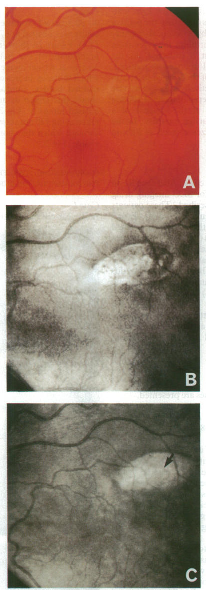
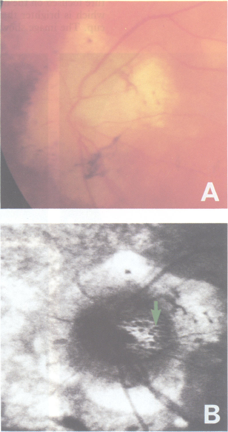
An infrared scanning laser ophthalmoscope (SLO) has been custom built in order to investigate the application of confocal and indirect mode SLO imaging to patients with fundus disease. Infrared light is reflected from the fundus to a greater extent than visible light permitting lower illumination power and, as it penetrates the retinal pigment epithelium, choroidal structures can be readily imaged. Furthermore, as conventional infrared illumination and detection systems are not well suited to ophthalmoscopy, this area is underdeveloped as a potential source of useful clinical data. Confocal, direct and indirect imaging modes have been used to image fundi of normal volunteers and patients with fundus disease. In comparison with conventional fundus photography confocal infrared SLO imaging improves visualisation of choroidal vasculature, retinal pigment epithelial abnormalities, laser photocoagulation scars, and optic disc pores in the lamina cribrosa. Direct infrared SLO imaging enables fundus visualisation through nuclear lens opacities. Furthermore, indirect mode imaging enhances significantly the appearance of macular drusen. The potential clinical benefit of these observations is discussed.

摘要

为了研究共聚焦和间接模式扫描激光检眼镜(SLO)成像在眼底疾病患者中的应用,我们特制了一台红外扫描激光检眼镜。红外光从眼底反射的程度比可见光更大,这使得照明功率更低,并且由于它能穿透视网膜色素上皮,脉络膜结构能够很容易地成像。此外,由于传统的红外照明和检测系统不太适合检眼镜检查,作为有用临床数据的潜在来源,这一领域尚未得到充分发展。共聚焦、直接和间接成像模式已被用于对正常志愿者和眼底疾病患者的眼底进行成像。与传统眼底摄影相比,共聚焦红外SLO成像改善了脉络膜血管系统、视网膜色素上皮异常、激光光凝瘢痕以及筛板层视盘孔的可视化。直接红外SLO成像能够透过核性晶状体混浊实现眼底可视化。此外,间接模式成像显著增强了黄斑玻璃膜疣的外观。我们还讨论了这些观察结果潜在的临床益处。

https://cdn.ncbi.nlm.nih.gov/pmc/blobs/ca66/504706/a5c522d6e842/brjopthal00026-0009-a.jpg
https://cdn.ncbi.nlm.nih.gov/pmc/blobs/ca66/504706/2086fbf98b72/brjopthal00026-0005-a.jpg
https://cdn.ncbi.nlm.nih.gov/pmc/blobs/ca66/504706/340dd34483de/brjopthal00026-0006-a.jpg
https://cdn.ncbi.nlm.nih.gov/pmc/blobs/ca66/504706/e4470136625a/brjopthal00026-0006-b.jpg
https://cdn.ncbi.nlm.nih.gov/pmc/blobs/ca66/504706/6e1fa2367225/brjopthal00026-0007-a.jpg
https://cdn.ncbi.nlm.nih.gov/pmc/blobs/ca66/504706/7e1ae6e19d4e/brjopthal00026-0007-b.jpg
https://cdn.ncbi.nlm.nih.gov/pmc/blobs/ca66/504706/8696a5d6f093/brjopthal00026-0007-c.jpg
https://cdn.ncbi.nlm.nih.gov/pmc/blobs/ca66/504706/438cdfd1568a/brjopthal00026-0008-a.jpg
https://cdn.ncbi.nlm.nih.gov/pmc/blobs/ca66/504706/411a35cbff78/brjopthal00026-0008-b.jpg
https://cdn.ncbi.nlm.nih.gov/pmc/blobs/ca66/504706/f7845966a258/brjopthal00026-0008-c.jpg
https://cdn.ncbi.nlm.nih.gov/pmc/blobs/ca66/504706/a5c522d6e842/brjopthal00026-0009-a.jpg

相似文献

1
Clinical investigation of an infrared digital scanning laser ophthalmoscope.一种红外数字扫描激光眼科镜的临床研究。
Br J Ophthalmol. 1994 Feb;78(2):84-90. doi: 10.1136/bjo.78.2.84.
2
Novel approach towards colour imaging using a scanning laser ophthalmoscope.使用扫描激光检眼镜进行彩色成像的新方法。
Br J Ophthalmol. 1998 Apr;82(4):342-5. doi: 10.1136/bjo.82.4.342.
3
Clinical investigation of a true color scanning laser ophthalmoscope.一种真彩色扫描激光检眼镜的临床研究。
Arch Ophthalmol. 2001 Jun;119(6):819-24. doi: 10.1001/archopht.119.6.819.
4
Confocal imaging of the fundus using a scanning laser ophthalmoscope.使用扫描激光检眼镜对眼底进行共聚焦成像。
Br J Ophthalmol. 1992 Aug;76(8):470-4. doi: 10.1136/bjo.76.8.470.
5
Fundus imaging in patients with cataract: role for a variable wavelength scanning laser ophthalmoscope.白内障患者的眼底成像:可变波长扫描激光检眼镜的作用
Br J Ophthalmol. 1995 Oct;79(10):892-9. doi: 10.1136/bjo.79.10.892.
6
Confocal scanning infrared laser ophthalmoscopy for indocyanine green angiography.用于吲哚菁绿血管造影的共焦扫描红外激光眼科检查法。
Am J Ophthalmol. 1995 Nov;120(5):642-51. doi: 10.1016/s0002-9394(14)72211-1.
7
Digital imaging of central serous retinopathy using infrared illumination.
Ger J Ophthalmol. 1995 Jul;4(4):203-6.
8
[New examination methods for macular disorders--application of diagnosis and treatment].[黄斑疾病的新检查方法——诊断与治疗的应用]
Nippon Ganka Gakkai Zasshi. 2000 Dec;104(12):899-942.
9
Confocal fundus imaging with a scanning laser ophthalmoscope in eyes with cataract.使用扫描激光检眼镜对白内障患者眼睛进行共焦眼底成像。
Br J Ophthalmol. 1995 Oct;79(10):900-4. doi: 10.1136/bjo.79.10.900.
10
Indocyanine green angiograms of choroidal nevi. comparison between confocal and nonconfocal scanning laser ophthalmoscope and fundus video camera.脉络膜痣的吲哚菁绿血管造影:共焦与非共焦扫描激光检眼镜及眼底摄像机的比较
Jpn J Ophthalmol. 2001 Jul-Aug;45(4):368-74. doi: 10.1016/s0021-5155(01)00362-8.

引用本文的文献

1
Infrared Photography: A Novel Diagnostic Approach for Ocular Surface Abnormalities Due to Vitamin A Deficiency.红外摄影:一种用于诊断维生素A缺乏所致眼表异常的新型方法。
Diagnostics (Basel). 2025 Jul 30;15(15):1910. doi: 10.3390/diagnostics15151910.
2
Retro-mode: a newer insight into dry age-related macular degeneration (AMD).返祖模式:干性年龄相关性黄斑变性(AMD)的新见解。
Lasers Med Sci. 2023 Aug 8;38(1):178. doi: 10.1007/s10103-023-03845-8.
3
Scanning laser ophthalmoscopy retroillumination: applications and illusions.扫描激光检眼镜后照法:应用与错觉

本文引用的文献

1
Digital fundus imaging using a scanning laser ophthalmoscope.
Physiol Meas. 1993 Feb;14(1):43-56. doi: 10.1088/0967-3334/14/1/006.
2
Fluorescein and indocyanine green angiographies of central serous choroidopathy by scanning laser ophthalmoscopy.通过扫描激光检眼镜进行的中心性浆液性脉络膜视网膜病变的荧光素和吲哚菁绿血管造影。
Am J Ophthalmol. 1993 Jan;115(1):50-6. doi: 10.1016/s0002-9394(14)73524-x.
3
Light levels in fundus photography and fluorescein angiography.
Vision Res. 1980;20(12):1099-104. doi: 10.1016/0042-6989(80)90046-2.
4
Int J Retina Vitreous. 2022 Sep 30;8(1):71. doi: 10.1186/s40942-022-00421-0.
4
Modern technologies for retinal scanning and imaging: an introduction for the biomedical engineer.视网膜扫描与成像的现代技术:给生物医学工程师的介绍
Biomed Eng Online. 2014 Apr 29;13:52. doi: 10.1186/1475-925X-13-52.
5
Optimization of confocal scanning laser ophthalmoscope design.共焦扫描激光检眼镜的设计优化。
J Biomed Opt. 2013 Jul;18(7):076015. doi: 10.1117/1.JBO.18.7.076015.
6
Infrared scanning laser ophthalmoscope imaging of the macula and its correlation with functional loss and structural changes in patients with stargardt disease.黄斑的红外扫描激光检眼镜成像及其与斯塔加特病患者的功能丧失和结构变化的相关性。
Retina. 2011 May;31(5):949-58. doi: 10.1097/IAE.0b013e3181f441f6.
7
Fine retinal crystalline deposits observed by confocal scanning laser ophthalmoscopic examination using infrared light.使用红外光的共焦扫描激光检眼镜检查观察到的视网膜细小晶体沉积物。
Br J Ophthalmol. 2003 Apr;87(4):509-10. doi: 10.1136/bjo.87.4.509.
8
Imaging of optic nerve head drusen with the scanning laser ophthalmoscope.使用扫描激光检眼镜对视乳头 drusen 进行成像。
Br J Ophthalmol. 1997 Aug;81(8):654-7. doi: 10.1136/bjo.81.8.654.
9
Observation of macular lesion in Best's vitelliform macular dystrophy by scanning laser ophthalmoscope.
Int Ophthalmol. 1995;19(6):369-73. doi: 10.1007/BF00130857.
10
Confocal fundus imaging with a scanning laser ophthalmoscope in eyes with cataract.使用扫描激光检眼镜对白内障患者眼睛进行共焦眼底成像。
Br J Ophthalmol. 1995 Oct;79(10):900-4. doi: 10.1136/bjo.79.10.900.
Council Lecture. Drusen and their relationship to senile macular degeneration.理事会讲座。玻璃膜疣及其与老年性黄斑变性的关系。
Aust J Ophthalmol. 1980 May;8(2):117-30. doi: 10.1111/j.1442-9071.1980.tb01670.x.
5
Ocular spectral characteristics as related to hazards from lasers and other light sources.
Am J Ophthalmol. 1968 Jul;66(1):15-20. doi: 10.1016/0002-9394(68)91780-7.
6
Color infrared photography of the ocular fundus.
Arch Ophthalmol. 1974 Sep;92(3):254-8. doi: 10.1001/archopht.1974.01010010262020.
7
Laser scanning: a method of retinal imaging.
Australas Phys Eng Sci Med. 1986 Oct-Dec;9(4):153-62.
8
The clinical appearance of the lamina cribrosa as a function of the extent of glaucomatous optic nerve damage.筛板的临床表现与青光眼性视神经损伤程度的关系。
Ophthalmology. 1988 Jan;95(1):135-8. doi: 10.1016/s0161-6420(88)33219-7.
9
Recording pattern reversal visual evoked response with the scanning laser ophthalmoscope.
Acta Ophthalmol (Copenh). 1989 Jun;67(3):243-8. doi: 10.1111/j.1755-3768.1989.tb01865.x.
10
Imaging retinal densitometry with a confocal Scanning Laser Ophthalmoscope.使用共焦扫描激光检眼镜进行视网膜密度成像。
Vision Res. 1989;29(12):1825-30. doi: 10.1016/0042-6989(89)90163-6.