Koski H, Konttinen Y T, Gu X H, Hietanen J, Malmström M
Department of Anatomy, University of Helsinki, Finland.
Ann Rheum Dis. 1995 Sep;54(9):744-7. doi: 10.1136/ard.54.9.744.
To compare the distribution and the amount of transforming growth factor beta (TGF beta) in labial salivary glands (LSG) in patients with Sjögren's syndrome (SS) and healthy controls.
LSG from SS patients (n = 10) and healthy controls (n = 6) were labelled with peroxidase-antiperoxidase staining for TGF beta 2, which was quantitated in image analysis using Video Interactive Display System software.
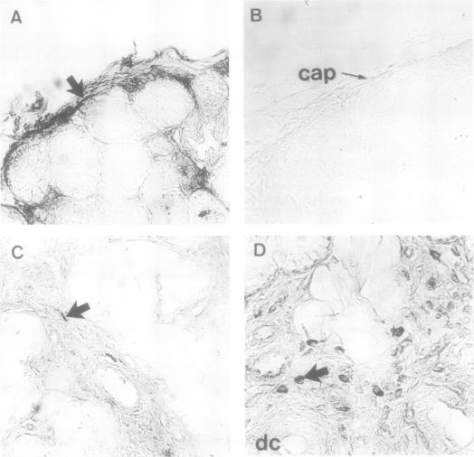
In all LSGs in SS and healthy controls, TGF beta 2 was found in endothelial cells of the capillaries and in the capsular and stromal fibroblasts. In LSGs in SS, TGF beta 2 was also found in some lymphocytes in the inflammatory cell foci and in fibroblasts in fibrotic areas. The TGF beta 2 staining index (microns 2/mm2 tissue) was greater in SS than in control LSGs (3670 (SEM 430) v 2061 (176); p < 0.01), with no difference between the primary and secondary forms of SS (p > 0.05).
The localisation and the level of expression of TGF beta 2 indicate its involvement in local tissue fibrosis, and may reflect attempts at immunosuppression.
比较干燥综合征(SS)患者和健康对照者唇腺(LSG)中转化生长因子β(TGFβ)的分布及含量。
对10例SS患者和6例健康对照者的LSG进行TGFβ2的过氧化物酶 - 抗过氧化物酶染色,使用视频交互式显示系统软件在图像分析中对其进行定量。
在SS患者和健康对照者的所有LSG中,TGFβ2存在于毛细血管内皮细胞、被膜和间质成纤维细胞中。在SS患者的LSG中,TGFβ2还存在于炎症细胞灶中的一些淋巴细胞以及纤维化区域的成纤维细胞中。SS患者LSG的TGFβ2染色指数(μm²/mm²组织)高于对照LSG(3670(标准误430)对2061(176);p < 0.01),SS的原发性和继发性形式之间无差异(p > 0.05)。
TGFβ2的定位和表达水平表明其参与局部组织纤维化,并可能反映免疫抑制的尝试。