Noteborn M H, Todd D, Verschueren C A, de Gauw H W, Curran W L, Veldkamp S, Douglas A J, McNulty M S, van der EB A J, Koch G
Laboratory for Molecular Carcinogenesis, Sylvius Laboratory, Leiden University, The Netherlands.
J Virol. 1994 Jan;68(1):346-51. doi: 10.1128/JVI.68.1.346-351.1994.
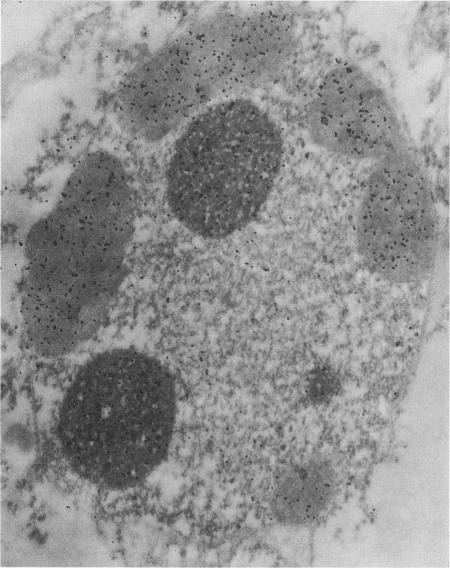
Chicken anemia virus (CAV) causes cytopathogenic effects in chicken thymocytes and cultured transformed mononuclear cells via apoptosis. Early after infection of chicken mononuclear cells, the CAV-encoded protein VP3 exhibits a finely granular distribution within the nucleus. At a later stage after infection, VP3 forms aggregates. At this point, the cell becomes apoptotic and the cellular DNA is fragmented and condensed. By immunogold electron microscopy VP3 was shown to be associated with apoptotic structures. In vitro, expression of VP3 induced apoptosis in chicken lymphoblastoid T cells and myeloid cells, which are susceptible to CAV infection, but not in chicken embryo fibroblasts, which are not susceptible to CAV. Expression of a C-terminally truncated VP3 induced much less pronounced apoptosis in the chicken lymphoblastoid T cells.
鸡贫血病毒(CAV)通过凋亡作用,在鸡胸腺细胞和培养的转化单核细胞中引起细胞病变效应。鸡单核细胞感染CAV后早期,CAV编码的蛋白VP3在细胞核内呈现精细的颗粒状分布。感染后期,VP3形成聚集体。此时,细胞发生凋亡,细胞DNA片段化并浓缩。通过免疫金电子显微镜观察发现,VP3与凋亡结构相关。在体外,VP3的表达在易受CAV感染的鸡淋巴母细胞样T细胞和髓样细胞中诱导凋亡,但在不易受CAV感染的鸡胚成纤维细胞中则不诱导凋亡。C末端截短的VP3在鸡淋巴母细胞样T细胞中诱导的凋亡明显较弱。