Kusumi A, Sako Y, Yamamoto M
Department of Pure and Applied Sciences, University of Tokyo, Japan.
Biophys J. 1993 Nov;65(5):2021-40. doi: 10.1016/S0006-3495(93)81253-0.
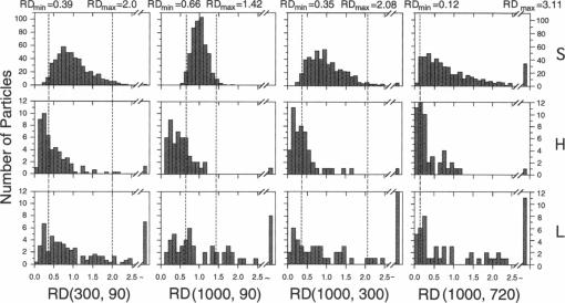
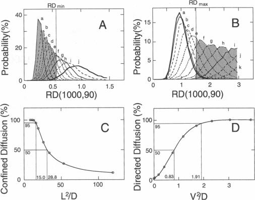
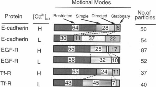
The movements of E-cadherin, epidermal growth factor receptor, and transferrin receptor in the plasma membrane of a cultured mouse keratinocyte cell line were studied using both single particle tracking (SPT; nanovid microscopy) and fluorescence photobleaching recovery (FPR). In the SPT technique, the receptor molecules are labeled with 40 nm-phi colloidal gold particles, and their movements are followed by video-enhanced differential interference contrast microscopy at a temporal resolution of 33 ms and at a nanometer-level spatial precision. The trajectories of the receptor molecules obtained by SPT were analyzed by developing a method that is based on the plot of the mean-square displacement against time. Four characteristic types of motion were observed: (a) stationary mode, in which the microscopic diffusion coefficient is less than 4.6 x 10(-12) cm2/s; (b) simple Brownian diffusion mode; (c) directed diffusion mode, in which unidirectional movements are superimposed on random motion; and (d) confined diffusion mode, in which particles undergoing Brownian diffusion (microscopic diffusion coefficient between 4.6 x 10(-12) and 1 x 10(-9) cm2/s) are confined within a limited area, probably by the membrane-associated cytoskeleton network. Comparison of these data obtained by SPT with those obtained by FPR suggests that the plasma membrane is compartmentalized into many small domains 300-600 nm in diameter (0.04-0.24 microns2 in area), in which receptor molecules are confined in the time scale of 3-30 s, and that the long-range diffusion observed by FPR can occur by successive movements of the receptors to adjacent compartments. Calcium-induced differentiation decreases the sum of the percentages of molecules in the directed diffusion and the stationary modes outside of the cell-cell contact regions on the cell surface (which is proposed to be the percentage of E-cadherin bound to the cytoskeleton/membrane-skeleton), from approximately 60% to 8% (low- and high-calcium mediums, respectively).
运用单粒子追踪技术(SPT;纳米视频显微镜)和荧光漂白恢复技术(FPR),对培养的小鼠角质形成细胞系质膜中E-钙黏蛋白、表皮生长因子受体和转铁蛋白受体的运动进行了研究。在SPT技术中,受体分子用40纳米直径的胶体金颗粒标记,通过视频增强微分干涉对比显微镜以33毫秒的时间分辨率和纳米级的空间精度追踪其运动。通过开发一种基于均方位移与时间关系图的方法,对SPT获得的受体分子轨迹进行了分析。观察到四种特征性的运动类型:(a)静止模式,其中微观扩散系数小于4.6×10⁻¹²平方厘米/秒;(b)简单布朗扩散模式;(c)定向扩散模式,其中单向运动叠加在随机运动上;(d)受限扩散模式,其中进行布朗扩散的颗粒(微观扩散系数在4.6×10⁻¹²至1×10⁻⁹平方厘米/秒之间)被限制在一个有限区域内,可能是由膜相关的细胞骨架网络所致。将SPT获得的数据与FPR获得的数据进行比较表明,质膜被分隔成许多直径为300 - 600纳米(面积为0.04 - 0.24微米²)的小区域,其中受体分子在3 - 30秒的时间尺度内被限制在这些区域,并且FPR观察到的长程扩散可能是由于受体向相邻区域的连续移动而发生的。钙诱导分化使细胞表面细胞 - 细胞接触区域之外的定向扩散和静止模式下分子的百分比总和(据推测这是与细胞骨架/膜骨架结合的E - 钙黏蛋白的百分比)从大约60%降至8%(分别为低钙和高钙培养基)。