• 文献检索
  • 文档翻译
  • 深度研究
  • 学术资讯
  • Suppr Zotero 插件Zotero 插件
  • 邀请有礼
  • 套餐&价格
  • 历史记录
应用&插件
Suppr Zotero 插件Zotero 插件浏览器插件Mac 客户端Windows 客户端微信小程序
定价
高级版会员购买积分包购买API积分包
服务
文献检索文档翻译深度研究API 文档MCP 服务
关于我们
关于 Suppr公司介绍联系我们用户协议隐私条款
关注我们

Suppr 超能文献

核心技术专利:CN118964589B侵权必究
粤ICP备2023148730 号-1Suppr @ 2026

文献检索

告别复杂PubMed语法,用中文像聊天一样搜索,搜遍4000万医学文献。AI智能推荐,让科研检索更轻松。

立即免费搜索

文件翻译

保留排版,准确专业,支持PDF/Word/PPT等文件格式,支持 12+语言互译。

免费翻译文档

深度研究

AI帮你快速写综述,25分钟生成高质量综述,智能提取关键信息,辅助科研写作。

立即免费体验

小鼠巨噬细胞动态产生肿瘤坏死因子-α(TNF-α)信使核糖核酸、细胞内和细胞外TNF-α以及与信号转导和转录激活因子1α(STAT1α)和细胞外信号调节激酶2(ERK2)蛋白酪氨酸磷酸化作为早期信号的可能关联。

Dynamic production of tumour necrosis factor-alpha (TNF-alpha) messenger RNA, intracellular and extracellular TNF-alpha by murine macrophages and possible association with protein tyrosine phosphorylation of STAT1 alpha and ERK2 as an early signal.

作者信息

Zheng Z M, Specter S

机构信息

Department of Medical Microbiology and Immunology, University of South Florida College of Medicine, FL, USA.

出版信息

Immunology. 1996 Apr;87(4):544-50. doi: 10.1046/j.1365-2567.1996.513591.x.

DOI:10.1046/j.1365-2567.1996.513591.x
PMID:8675207
原文链接:https://pmc.ncbi.nlm.nih.gov/articles/PMC1384131/
Abstract

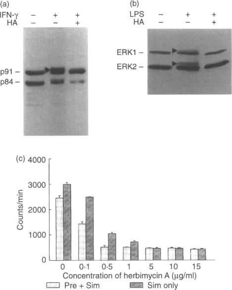
Tumour necrosis factor-alpha (TNF-alpha), an important mediator in both immune and inflammation responses, is one of the major cytokines released by activated macrophages. The present study shows that, during macrophage activation, protein tyrosine phosphorylation of STAT1 alpha and ERK2 occurred as an immediate early signal, whereas maximum TNF-alpha mRNA transcription appeared at 3 hr, precursor TNF-alpha formation at 3 to 4 hr, and TNF-alpha release at 5 to 6 hr after stimulation of an RPMI-1640-based induction medium containing lipopolysaccharide (100 ng/ml), interferon-gamma (100 U/ml), and 0.5% bovine serum albumin. Herbimycin A, a tyrosine kinase inhibitor, suppresses protein tyrosine phosphorylation of STAT1 alpha and ERK2 and also blocks TNF-alpha production by resident peritoneal macrophages from BALB/c mice, suggesting a possible association between protein tyrosine phosphorylation of STAT1 alpha and ERK2 and macrophage activation resulting in TNF-alpha production.

摘要

肿瘤坏死因子-α(TNF-α)是免疫和炎症反应中的一种重要介质,是活化巨噬细胞释放的主要细胞因子之一。本研究表明,在巨噬细胞活化过程中,STAT1α和ERK2的蛋白酪氨酸磷酸化作为即时早期信号出现,而在含有脂多糖(100 ng/ml)、干扰素-γ(100 U/ml)和0.5%牛血清白蛋白的基于RPMI-1640的诱导培养基刺激后,TNF-α mRNA转录在3小时达到最大值,前体TNF-α在3至4小时形成,TNF-α在5至6小时释放。酪氨酸激酶抑制剂赫比霉素A可抑制STAT1α和ERK2的蛋白酪氨酸磷酸化,也可阻断BALB/c小鼠腹腔驻留巨噬细胞产生TNF-α,这表明STAT1α和ERK2的蛋白酪氨酸磷酸化与巨噬细胞活化导致TNF-α产生之间可能存在关联。

https://cdn.ncbi.nlm.nih.gov/pmc/blobs/4a89/1384131/9f3b7dff36fc/immunology00061-0038-b.jpg
https://cdn.ncbi.nlm.nih.gov/pmc/blobs/4a89/1384131/b87ecf3da96e/immunology00061-0036-a.jpg
https://cdn.ncbi.nlm.nih.gov/pmc/blobs/4a89/1384131/513ad0f87065/immunology00061-0036-b.jpg
https://cdn.ncbi.nlm.nih.gov/pmc/blobs/4a89/1384131/d9db8b4bc8a5/immunology00061-0037-a.jpg
https://cdn.ncbi.nlm.nih.gov/pmc/blobs/4a89/1384131/90277e2813b7/immunology00061-0038-a.jpg
https://cdn.ncbi.nlm.nih.gov/pmc/blobs/4a89/1384131/9f3b7dff36fc/immunology00061-0038-b.jpg

相似文献

1
Dynamic production of tumour necrosis factor-alpha (TNF-alpha) messenger RNA, intracellular and extracellular TNF-alpha by murine macrophages and possible association with protein tyrosine phosphorylation of STAT1 alpha and ERK2 as an early signal.小鼠巨噬细胞动态产生肿瘤坏死因子-α(TNF-α)信使核糖核酸、细胞内和细胞外TNF-α以及与信号转导和转录激活因子1α(STAT1α)和细胞外信号调节激酶2(ERK2)蛋白酪氨酸磷酸化作为早期信号的可能关联。
Immunology. 1996 Apr;87(4):544-50. doi: 10.1046/j.1365-2567.1996.513591.x.
2
Modulation of TNF-alpha gene expression by IFN-gamma and pamidronate in murine macrophages: regulation by STAT1-dependent pathways.
J Immunol. 2005 Feb 15;174(4):1801-10. doi: 10.4049/jimmunol.174.4.1801.
3
Activation of Raf-1 and mitogen-activated protein kinase in murine macrophages partially mimics lipopolysaccharide-induced signaling events.鼠巨噬细胞中Raf-1和丝裂原活化蛋白激酶的激活部分模拟了脂多糖诱导的信号事件。
J Exp Med. 1995 Jul 1;182(1):147-54. doi: 10.1084/jem.182.1.147.
4
Mycobacterium avium infection of mouse macrophages inhibits IFN-gamma Janus kinase-STAT signaling and gene induction by down-regulation of the IFN-gamma receptor.鸟分枝杆菌对小鼠巨噬细胞的感染通过下调γ干扰素受体来抑制γ干扰素 Janus 激酶-信号转导和转录激活因子信号传导及基因诱导。
J Immunol. 1999 Aug 15;163(4):2041-8.
5
Bovine serum albumin preparations enhance in vitro production of tumor necrosis factor alpha by murine macrophages.牛血清白蛋白制剂可增强小鼠巨噬细胞体外产生肿瘤坏死因子α的能力。
Immunol Invest. 1995 Aug;24(5):737-56. doi: 10.3109/08820139509060702.
6
In vitro activation of murine peritoneal macrophages by monocyte chemoattractant protein-1: upregulation of CD11b, production of proinflammatory cytokines, and the signal transduction pathway.单核细胞趋化蛋白-1对小鼠腹腔巨噬细胞的体外激活:CD11b的上调、促炎细胞因子的产生及信号转导途径
J Interferon Cytokine Res. 2002 May;22(5):527-38. doi: 10.1089/10799900252982007.
7
Lipopolysaccharide signals activation of tumor necrosis factor biosynthesis through the ras/raf-1/MEK/MAPK pathway.脂多糖通过ras/raf-1/MEK/MAPK途径激活肿瘤坏死因子的生物合成。
Mol Med. 1994 Nov;1(1):93-103.
8
Preferential involvement of MEK1 in the tumor necrosis factor-alpha-induced activation of p42mapk/erk2 in mouse macrophages.MEK1在肿瘤坏死因子-α诱导的小鼠巨噬细胞中p42mapk/erk2激活过程中的优先参与。
J Biol Chem. 1995 Nov 17;270(46):27391-4. doi: 10.1074/jbc.270.46.27391.
9
Stress-induced phosphorylation of STAT1 at Ser727 requires p38 mitogen-activated protein kinase whereas IFN-gamma uses a different signaling pathway.应激诱导的信号转导和转录激活因子1(STAT1)在丝氨酸727位点的磷酸化需要p38丝裂原活化蛋白激酶,而γ干扰素则使用不同的信号通路。
Proc Natl Acad Sci U S A. 1999 Nov 23;96(24):13956-61. doi: 10.1073/pnas.96.24.13956.
10
Activation of p42mapk/erk2 following engagement of tumor necrosis factor receptor CD120a (p55) in mouse macrophages.在小鼠巨噬细胞中,肿瘤坏死因子受体CD120a(p55)被激活后p42mapk/erk2的激活。
J Immunol. 1995 Aug 1;155(3):1525-33.

引用本文的文献

1
STAT1 inhibits T-cell exhaustion and myeloid derived suppressor cell accumulation to promote antitumor immune responses in head and neck squamous cell carcinoma.STAT1 抑制 T 细胞耗竭和髓系来源的抑制细胞积累,以促进头颈部鳞状细胞癌中的抗肿瘤免疫反应。
Int J Cancer. 2020 Mar 15;146(6):1717-1729. doi: 10.1002/ijc.32781. Epub 2019 Nov 29.
2
The equine alveolar macrophage: functional and phenotypic comparisons with peritoneal macrophages.马肺泡巨噬细胞:与腹腔巨噬细胞的功能和表型比较
Vet Immunol Immunopathol. 2013 Oct 1;155(4):219-28. doi: 10.1016/j.vetimm.2013.07.003. Epub 2013 Jul 20.
3
Emerging roles for TNIP1 in regulating post-receptor signaling.

本文引用的文献

1
Bovine serum albumin preparations enhance in vitro production of tumor necrosis factor alpha by murine macrophages.牛血清白蛋白制剂可增强小鼠巨噬细胞体外产生肿瘤坏死因子α的能力。
Immunol Invest. 1995 Aug;24(5):737-56. doi: 10.3109/08820139509060702.
2
Signal transduction via the MAP kinases: proceed at your own RSK.通过丝裂原活化蛋白激酶的信号转导:自行承担风险进行。 (注:“proceed at your own RSK”表述不太常规,可能在特定语境中有特殊含义,这里按字面大致翻译,其中“RSK”可能是特定术语首字母缩写,不太明确其准确含义)
Proc Natl Acad Sci U S A. 1993 Jul 1;90(13):5889-92. doi: 10.1073/pnas.90.13.5889.
3
Tyrosine phosphorylation of mitogen-activated protein kinases is necessary for activation of murine macrophages by natural and synthetic bacterial products.
TNIP1 在调节受体后信号转导中的新兴作用。
Cytokine Growth Factor Rev. 2012 Jun;23(3):109-18. doi: 10.1016/j.cytogfr.2012.04.002. Epub 2012 Apr 28.
4
Signals that dictate nuclear localization of human papillomavirus type 16 oncoprotein E6 in living cells.决定人乳头瘤病毒16型癌蛋白E6在活细胞中核定位的信号。
J Virol. 2003 Dec;77(24):13232-47. doi: 10.1128/jvi.77.24.13232-13247.2003.
5
Determination of optimal sites of antisense oligonucleotide cleavage within TNFalpha mRNA.肿瘤坏死因子α(TNFα)信使核糖核酸(mRNA)内反义寡核苷酸切割最佳位点的确定
Nucleic Acids Res. 2001 Sep 1;29(17):3664-73. doi: 10.1093/nar/29.17.3664.
6
The virulence plasmid of Yersinia, an antihost genome.耶尔森氏菌的毒力质粒,一种抗宿主基因组。
Microbiol Mol Biol Rev. 1998 Dec;62(4):1315-52. doi: 10.1128/MMBR.62.4.1315-1352.1998.
7
Role of YopP in suppression of tumor necrosis factor alpha release by macrophages during Yersinia infection.耶尔森氏菌感染期间YopP在抑制巨噬细胞释放肿瘤坏死因子α中的作用。
Infect Immun. 1998 May;66(5):1878-84. doi: 10.1128/IAI.66.5.1878-1884.1998.
8
Yersinia enterocolitica impairs activation of transcription factor NF-kappaB: involvement in the induction of programmed cell death and in the suppression of the macrophage tumor necrosis factor alpha production.小肠结肠炎耶尔森菌损害转录因子NF-κB的激活:参与程序性细胞死亡的诱导及巨噬细胞肿瘤坏死因子α产生的抑制。
J Exp Med. 1998 Apr 6;187(7):1069-79. doi: 10.1084/jem.187.7.1069.
丝裂原活化蛋白激酶的酪氨酸磷酸化是天然和合成细菌产物激活小鼠巨噬细胞所必需的。
J Exp Med. 1993 Apr 1;177(4):1071-7. doi: 10.1084/jem.177.4.1071.
4
Induction by EGF and interferon-gamma of tyrosine phosphorylated DNA binding proteins in mouse liver nuclei.表皮生长因子(EGF)和γ干扰素对小鼠肝细胞核中酪氨酸磷酸化DNA结合蛋白的诱导作用
Science. 1993 Sep 24;261(5129):1733-6. doi: 10.1126/science.8378774.
5
Differential expression of tumor necrosis factor and interleukin-6 by peritoneal macrophages in vivo and in culture.体内及培养环境中腹膜巨噬细胞对肿瘤坏死因子和白细胞介素-6的差异表达
Am J Pathol. 1993 Oct;143(4):1121-30.
6
Jak-STAT pathways and transcriptional activation in response to IFNs and other extracellular signaling proteins.Jak-STAT信号通路以及对干扰素和其他细胞外信号蛋白的转录激活。
Science. 1994 Jun 3;264(5164):1415-21. doi: 10.1126/science.8197455.
7
delta 9-Tetrahydrocannabinol (THC) modulates IL-1 bioactivity in human monocyte/macrophage cell lines.
Life Sci. 1994;54(17):1281-9. doi: 10.1016/0024-3205(94)00856-6.
8
Okadaic acid stimulates inflammatory cytokine gene transcription in murine peritoneal macrophages.冈田酸刺激小鼠腹腔巨噬细胞中炎性细胞因子基因转录。
Cell Immunol. 1994 Feb;153(2):479-91. doi: 10.1006/cimm.1994.1044.
9
Suppression by delta-9-tetrahydrocannabinol of lipopolysaccharide-induced and intrinsic tyrosine phosphorylation and protein expression in mouse peritoneal macrophages.δ-9-四氢大麻酚对小鼠腹腔巨噬细胞中脂多糖诱导的和内在的酪氨酸磷酸化及蛋白质表达的抑制作用。
Biochem Pharmacol. 1994 Jun 15;47(12):2243-52. doi: 10.1016/0006-2952(94)90262-3.
10
Transcriptional regulation by extracellular signals: mechanisms and specificity.
Cell. 1995 Jan 27;80(2):199-211. doi: 10.1016/0092-8674(95)90403-4.