• 文献检索
  • 文档翻译
  • 深度研究
  • 学术资讯
  • Suppr Zotero 插件Zotero 插件
  • 邀请有礼
  • 套餐&价格
  • 历史记录
应用&插件
Suppr Zotero 插件Zotero 插件浏览器插件Mac 客户端Windows 客户端微信小程序
定价
高级版会员购买积分包购买API积分包
服务
文献检索文档翻译深度研究API 文档MCP 服务
关于我们
关于 Suppr公司介绍联系我们用户协议隐私条款
关注我们

Suppr 超能文献

核心技术专利:CN118964589B侵权必究
粤ICP备2023148730 号-1Suppr @ 2026

文献检索

告别复杂PubMed语法,用中文像聊天一样搜索,搜遍4000万医学文献。AI智能推荐,让科研检索更轻松。

立即免费搜索

文件翻译

保留排版,准确专业,支持PDF/Word/PPT等文件格式,支持 12+语言互译。

免费翻译文档

深度研究

AI帮你快速写综述,25分钟生成高质量综述,智能提取关键信息,辅助科研写作。

立即免费体验

使用光学相干断层扫描技术测量神经纤维层厚度的可重复性。

Reproducibility of nerve fiber layer thickness measurements using optical coherence tomography.

作者信息

Schuman J S, Pedut-Kloizman T, Hertzmark E, Hee M R, Wilkins J R, Coker J G, Puliafito C A, Fujimoto J G, Swanson E A

机构信息

New England Eye Center, Tufts University School of Medicine, Boston, MA 02111, USA.

出版信息

Ophthalmology. 1996 Nov;103(11):1889-98. doi: 10.1016/s0161-6420(96)30410-7.

DOI:10.1016/s0161-6420(96)30410-7
PMID:8942887
原文链接:https://pmc.ncbi.nlm.nih.gov/articles/PMC1939724/
Abstract

PURPOSE

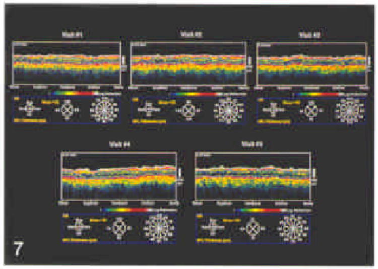
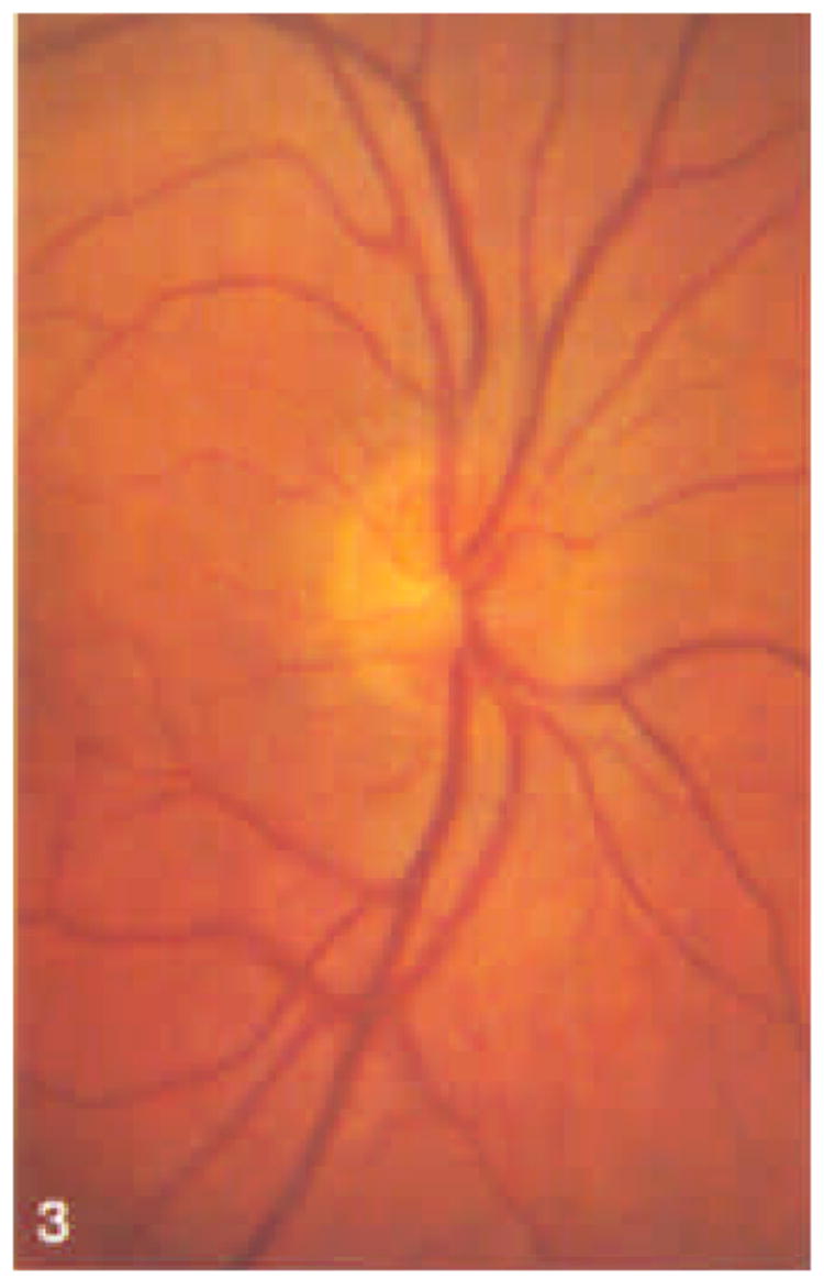
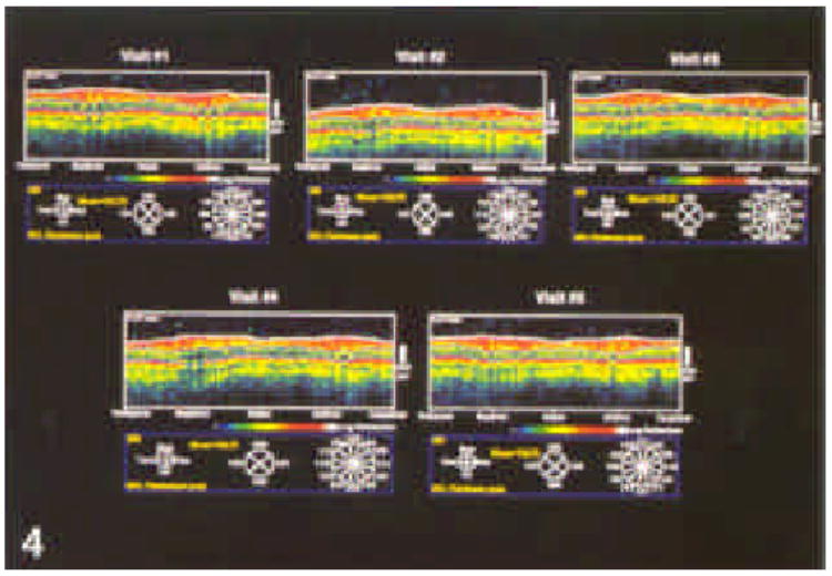
Optical coherence tomography (OCT) is a new technology that uses near-infrared light in an interferometer to produce approximately 10-microns resolution cross-sectional images of the tissue of interest. The authors performed repeated quantitative assessment of nerve fiber layer thickness in individuals with normal and glaucomatous eyes, and they evaluated the reproducibility of these measurements.

METHODS

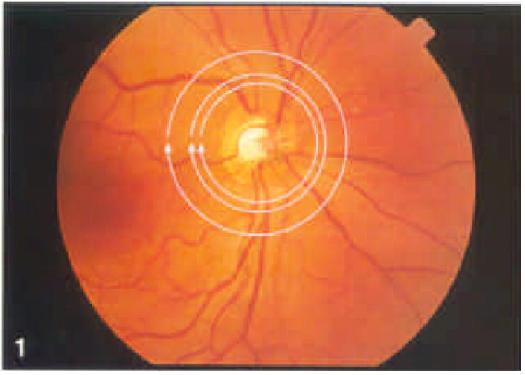
The authors studied 21 eyes of 21 subjects by OCT. Each subject underwent five repetitions of a series of scans on five separate occasions within a 1-month period. Each series consisted of three circular scans around the optic nerve head (diameters, 2.9, 3.4, and 4.5 mm). Each series was performed separately using internal (fixation with same eye being studied) and external (fixation with contralateral eye) fixation techniques. The eye studied and the sequence of testing were assigned randomly.

RESULTS

Internal fixation (IF), in general, provides a slightly higher degree of reproducibility than external fixation (EF). Reproducibility was better in a given eye on a given visit than from visit to visit. Reproducibility as measured by intraclass correlation coefficients were as follows: circle diameter (CD), 2.9 mm, 0.51/0.57 (normal/glaucoma) (IF), 0.43/0.54 (EF); CD, 3.4 mm, 0.56/0.52 (IF), 0.43/0.61 (EF); CD, 4.5 mm, 0.53/0.43 (IF), 0.42/0.49 (EF).

CONCLUSIONS

Nerve fiber layer thickness can be reproducibly measured using OCT. Internal is superior to external fixation; each circle diameter tested provides adequate reproducibility.

摘要

目的

光学相干断层扫描(OCT)是一项新技术,它在干涉仪中使用近红外光来生成感兴趣组织的约10微米分辨率的横截面图像。作者对正常眼和青光眼患者的神经纤维层厚度进行了重复定量评估,并评估了这些测量的可重复性。

方法

作者通过OCT研究了21名受试者的21只眼睛。每位受试者在1个月内的5个不同时间对一系列扫描进行了5次重复。每个系列包括围绕视神经乳头的三次圆形扫描(直径分别为2.9、3.4和4.5毫米)。每个系列分别使用内固定(用被研究的同一只眼睛注视)和外固定(用对侧眼睛注视)技术进行。被研究的眼睛和测试顺序是随机分配的。

结果

一般来说,内固定(IF)比外固定(EF)提供的可重复性略高。在给定的一次就诊中,给定眼睛的可重复性比不同就诊之间的更好。通过组内相关系数测量的可重复性如下:圆直径(CD)为2.9毫米时,0.51/0.57(正常/青光眼)(IF),0.43/0.54(EF);CD为3.4毫米时,0.56/0.52(IF),0.43/0.61(EF);CD为4.5毫米时,0.53/0.43(IF),0.42/0.49(EF)。

结论

使用OCT可以可重复地测量神经纤维层厚度。内固定优于外固定;所测试的每个圆直径都提供了足够的可重复性。

https://cdn.ncbi.nlm.nih.gov/pmc/blobs/d895/1939724/bd5ddc97230c/nihms24601f7.jpg
https://cdn.ncbi.nlm.nih.gov/pmc/blobs/d895/1939724/c60e6f22d585/nihms24601f1.jpg
https://cdn.ncbi.nlm.nih.gov/pmc/blobs/d895/1939724/60405878c033/nihms24601f2.jpg
https://cdn.ncbi.nlm.nih.gov/pmc/blobs/d895/1939724/113f37dc54aa/nihms24601f3.jpg
https://cdn.ncbi.nlm.nih.gov/pmc/blobs/d895/1939724/572f028375ed/nihms24601f4.jpg
https://cdn.ncbi.nlm.nih.gov/pmc/blobs/d895/1939724/879da8caf48b/nihms24601f5.jpg
https://cdn.ncbi.nlm.nih.gov/pmc/blobs/d895/1939724/41eb70031dde/nihms24601f6.jpg
https://cdn.ncbi.nlm.nih.gov/pmc/blobs/d895/1939724/bd5ddc97230c/nihms24601f7.jpg

相似文献

1
Reproducibility of nerve fiber layer thickness measurements using optical coherence tomography.使用光学相干断层扫描技术测量神经纤维层厚度的可重复性。
Ophthalmology. 1996 Nov;103(11):1889-98. doi: 10.1016/s0161-6420(96)30410-7.
2
Quantification of nerve fiber layer thickness in normal and glaucomatous eyes using optical coherence tomography.使用光学相干断层扫描技术对正常眼和青光眼眼中神经纤维层厚度进行定量分析。
Arch Ophthalmol. 1995 May;113(5):586-96. doi: 10.1001/archopht.1995.01100050054031.
3
Reproducibility of nerve fiber layer thickness measurements using optical coherence tomography in silicone oil-filled eyes.在硅油填充眼中使用光学相干断层扫描测量神经纤维层厚度的可重复性。
Ophthalmologica. 2001 Mar-Apr;215(2):91-6. doi: 10.1159/000050836.
4
Evaluation of focal defects of the nerve fiber layer using optical coherence tomography.使用光学相干断层扫描技术评估神经纤维层的局灶性缺损。
Ophthalmology. 1999 Mar;106(3):570-9. doi: 10.1016/s0161-6420(99)90118-5.
5
Reliability of nerve fiber layer thickness measurements using optical coherence tomography in normal and glaucomatous eyes.光学相干断层扫描测量正常眼和青光眼眼中神经纤维层厚度的可靠性。
Ophthalmology. 2003 Jan;110(1):190-5. doi: 10.1016/s0161-6420(02)01296-4.
6
Optical coherence tomography in measuring retinal nerve fiber layer thickness in normal subjects and patients with open-angle glaucoma.光学相干断层扫描技术在测量正常受试者和开角型青光眼患者视网膜神经纤维层厚度中的应用
Chin Med J (Engl). 2001 May;114(5):524-9.
7
Optical coherence tomography: a new tool for glaucoma diagnosis.光学相干断层扫描:青光眼诊断的新工具。
Curr Opin Ophthalmol. 1995 Apr;6(2):89-95. doi: 10.1097/00055735-199504000-00014.
8
Reproducibility of peripapillary retinal nerve fiber layer thickness measurements using spectral domain OCT in Brazilian patients.巴西患者中使用光谱域光学相干断层扫描测量视乳头周围视网膜神经纤维层厚度的可重复性。
Arq Bras Oftalmol. 2012 Oct;75(5):320-3. doi: 10.1590/s0004-27492012000500005.
9
Detection of Progressive Glaucomatous Optic Nerve Damage on Fundus Photographs with Deep Learning.利用深度学习技术在眼底照片上检测青光眼性视神经进行性损伤
Ophthalmology. 2021 Mar;128(3):383-392. doi: 10.1016/j.ophtha.2020.07.045. Epub 2020 Jul 28.
10
2-D pattern of nerve fiber bundles in glaucoma emerging from spectral-domain optical coherence tomography.青光眼的神经纤维束的二维模式从频域光学相干断层扫描中显现出来。
Invest Ophthalmol Vis Sci. 2012 Jan 31;53(1):483-9. doi: 10.1167/iovs.11-8349.

引用本文的文献

1
Novel Structure-Function Models for Estimating Retinal Ganglion Cell Count Using Pattern Electroretinography in Glaucoma Suspects.用于在青光眼疑似患者中使用图形视网膜电图估计视网膜神经节细胞数量的新型结构-功能模型。
Diagnostics (Basel). 2025 Jul 11;15(14):1756. doi: 10.3390/diagnostics15141756.
2
Retinal Imaging as a Window into Cardiovascular Health: Towards Harnessing Retinal Analytics for Precision Cardiovascular Medicine.视网膜成像作为洞察心血管健康的窗口:迈向利用视网膜分析实现精准心血管医学
J Cardiovasc Dev Dis. 2025 Jun 17;12(6):230. doi: 10.3390/jcdd12060230.
3
Spectralis Optical Coherence Tomography for Evaluating Ocular Hypertensive and Glaucoma Suspect Eyes: Real-World Data from Taiwan.

本文引用的文献

1
Repeatability of the glaucoma-scope measurements of optic nerve head topography.青光眼眼底仪视神经头地形图测量的重复性。
J Glaucoma. 1994 Spring;3(1):17-27.
2
In vivo retinal imaging by optical coherence tomography.通过光学相干断层扫描进行视网膜活体成像。
Opt Lett. 1993 Nov 1;18(21):1864-6. doi: 10.1364/ol.18.001864.
3
Effect of repetitive imaging on topographic measurements of the optic nerve head.重复成像对视神经乳头地形图测量的影响。
使用Spectralis光学相干断层扫描技术评估高眼压症和疑似青光眼患者的眼部:来自台湾的真实世界数据
Diagnostics (Basel). 2025 May 15;15(10):1256. doi: 10.3390/diagnostics15101256.
4
Performance of Optic Disc Optical Coherence Tomography Normative Database in a Large, Diverse, Real-World Cohort.视盘光学相干断层扫描标准数据库在一个大型、多样化的真实世界队列中的表现
J Glaucoma. 2025 May 1;34(5):404-414. doi: 10.1097/IJG.0000000000002545. Epub 2025 Jan 30.
5
Diagnostic Capability of Pattern Electroretinogram and Three Circumpapillary Retinal Nerve Fiber Layer Thickness Circle Diameter Scans in Glaucoma Suspects.图形视网膜电图及三次视盘周围视网膜神经纤维层厚度圆周直径扫描对青光眼可疑患者的诊断能力
Clin Ophthalmol. 2025 Jan 22;19:235-247. doi: 10.2147/OPTH.S483055. eCollection 2025.
6
Prediction of Visual Field Progression with Baseline and Longitudinal Structural Measurements Using Deep Learning.基于深度学习的基线和纵向结构测量对视功能进展的预测。
Am J Ophthalmol. 2024 Jun;262:141-152. doi: 10.1016/j.ajo.2024.02.007. Epub 2024 Feb 12.
7
How Radioactive Iodine Treatment Affects the Retina.放射性碘治疗如何影响视网膜。
World J Nucl Med. 2023 Dec 4;22(4):257-260. doi: 10.1055/s-0043-1774419. eCollection 2023 Dec.
8
Evaluation of Optical Coherence Tomography Findings in Antisocial Personality Disorder Patients.反社会型人格障碍患者的光学相干断层扫描结果评估
Alpha Psychiatry. 2023 Jul 1;24(4):121-127. doi: 10.5152/alphapsychiatry.2023.221108. eCollection 2023 Jul.
9
Artifact Correction in Retinal Nerve Fiber Layer Thickness Maps Using Deep Learning and Its Clinical Utility in Glaucoma.基于深度学习的视网膜神经纤维层厚度图的伪影校正及其在青光眼临床中的应用。
Transl Vis Sci Technol. 2023 Nov 1;12(11):12. doi: 10.1167/tvst.12.11.12.
10
Focused analysis of RNFL decay in glaucomatous eyes using circular statistics on high-resolution OCT data.利用高分辨率 OCT 数据的圆形统计学对青光眼眼的 RNFL 衰减进行集中分析。
PLoS One. 2023 Oct 18;18(10):e0292915. doi: 10.1371/journal.pone.0292915. eCollection 2023.
Arch Ophthalmol. 1993 May;111(5):636-8. doi: 10.1001/archopht.1993.01090050070031.
4
Micrometer-scale resolution imaging of the anterior eye in vivo with optical coherence tomography.利用光学相干断层扫描对活体眼前部进行微米级分辨率成像。
Arch Ophthalmol. 1994 Dec;112(12):1584-9. doi: 10.1001/archopht.1994.01090240090031.
5
Optical coherence tomography of the human retina.人类视网膜的光学相干断层扫描。
Arch Ophthalmol. 1995 Mar;113(3):325-32. doi: 10.1001/archopht.1995.01100030081025.
6
Imaging of macular diseases with optical coherence tomography.光学相干断层扫描对黄斑疾病的成像
Ophthalmology. 1995 Feb;102(2):217-29. doi: 10.1016/s0161-6420(95)31032-9.
7
Optical coherence tomography of macular holes.黄斑裂孔的光学相干断层扫描
Ophthalmology. 1995 May;102(5):748-56. doi: 10.1016/s0161-6420(95)30959-1.
8
Quantification of nerve fiber layer thickness in normal and glaucomatous eyes using optical coherence tomography.使用光学相干断层扫描技术对正常眼和青光眼眼中神经纤维层厚度进行定量分析。
Arch Ophthalmol. 1995 May;113(5):586-96. doi: 10.1001/archopht.1995.01100050054031.
9
Scanning laser polarimetry to measure the nerve fiber layer of normal and glaucomatous eyes.扫描激光偏振仪测量正常眼和青光眼患者的神经纤维层。
Am J Ophthalmol. 1995 May;119(5):627-36. doi: 10.1016/s0002-9394(14)70221-1.
10
Reproducibility of topographic measurements of the optic nerve head with laser tomographic scanning.
Ophthalmology. 1989 Sep;96(9):1320-4. doi: 10.1016/s0161-6420(89)32719-9.