D'Souza S D, Bonetti B, Balasingam V, Cashman N R, Barker P A, Troutt A B, Raine C S, Antel J P
Neuroimmunology Unit, McGill University, Montreal Neurological Institute, Quebec, Canada.
J Exp Med. 1996 Dec 1;184(6):2361-70. doi: 10.1084/jem.184.6.2361.
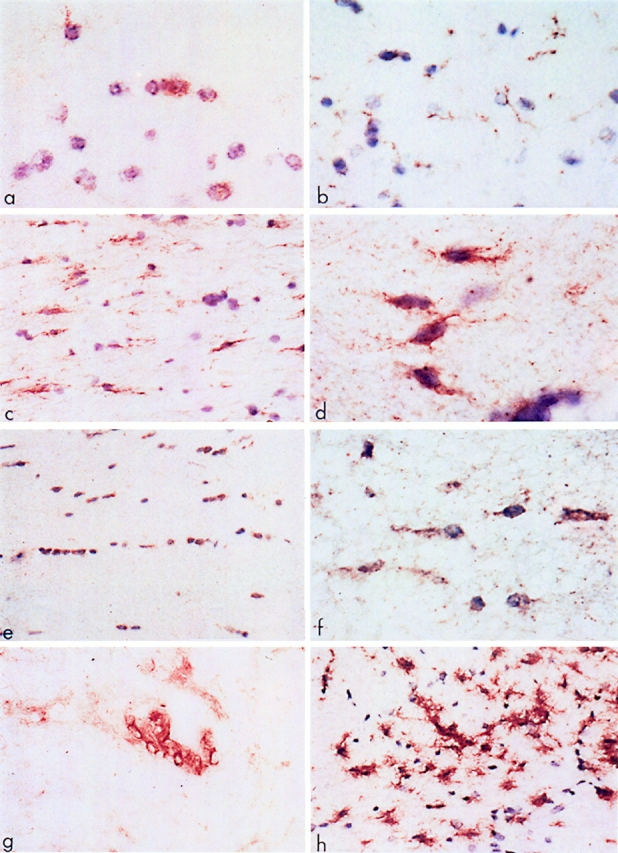
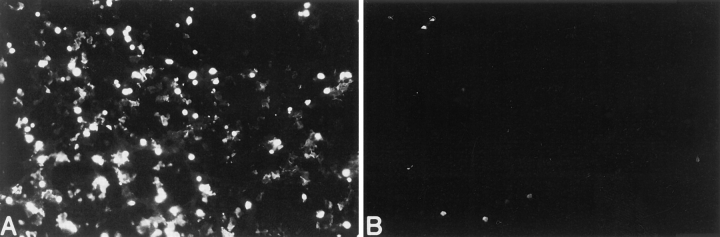
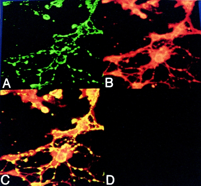
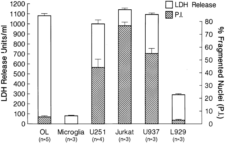
Fas is a cell surface receptor that transduces cell death signals when cross-linked by agonist antibodies or by fas ligand. In this study, we examined the potential of fas to contribute to oligodendrocyte (OL) injury and demyelination as they occur in the human demyelinating disease multiple sclerosis (MS). Immunohistochemical study of central nervous system (CNS) tissue from MS subjects demonstrated elevated fas expression on OLs in chronic active and chronic silent MS lesions compared with OLs in control tissue from subjects with or without other neurologic diseases. In such lesions, microglia and infiltrating lymphocytes displayed intense immunoreactivity to fas ligand. In dissociated glial cell cultures prepared from human adult CNS tissue, fas expression was restricted to OLs. Fas ligation with the anti-fas monoclonal antibody M3 or with the fas-ligand induced rapid OL cell membrane lysis, assessed by LDH release and trypan blue uptake and subsequent cell death. In contrast to the activity of fas in other cellular systems, dying OLs did not exhibit evidence of apoptosis, assessed morphologically and by terminal transferase-mediated d-uridine triphosphate-biotin nick-end-labeling staining for DNA fragmentation. Other stimuli such as C2-ceramide were capable of inducing rapid apoptosis in OLs. Antibodies directed at other surface molecules expressed on OLs or the M33 non-activating anti-fas monoclonal antibody did not induce cytolysis of OLs. Our results suggest that fas-mediated signaling might contribute in a novel cytolytic manner to immune-mediated OL injury in MS.
Fas是一种细胞表面受体,当被激动剂抗体或Fas配体交联时可转导细胞死亡信号。在本研究中,我们探讨了Fas在人类脱髓鞘疾病多发性硬化症(MS)中导致少突胶质细胞(OL)损伤和脱髓鞘的可能性。对MS患者中枢神经系统(CNS)组织的免疫组织化学研究表明,与患有或未患有其他神经系统疾病的对照组织中的OL相比,慢性活动期和慢性静止期MS病变中的OL上Fas表达升高。在这些病变中,小胶质细胞和浸润淋巴细胞对Fas配体表现出强烈的免疫反应性。在从成人人类CNS组织制备的解离神经胶质细胞培养物中,Fas表达仅限于OL。用抗Fas单克隆抗体M3或Fas配体连接Fas可诱导快速的OL细胞膜裂解,通过乳酸脱氢酶(LDH)释放、台盼蓝摄取及随后的细胞死亡进行评估。与Fas在其他细胞系统中的活性不同,垂死的OL未表现出凋亡迹象,通过形态学评估以及用于DNA片段化的末端转移酶介导的d-三磷酸尿苷-生物素缺口末端标记染色评估。其他刺激物如C2-神经酰胺能够诱导OL快速凋亡。针对OL上表达的其他表面分子的抗体或M33非激活抗Fas单克隆抗体不会诱导OL的细胞溶解。我们的结果表明,Fas介导的信号传导可能以一种新的细胞溶解方式促成MS中免疫介导的OL损伤。