King J M, Hays T S, Nicklas R B
Department of Biology, Duke University, Durham, North Carolina 27708, USA.
J Cell Biol. 2000 Nov 13;151(4):739-48. doi: 10.1083/jcb.151.4.739.
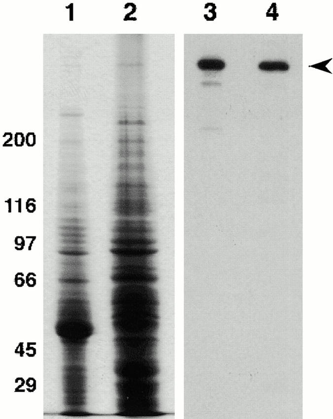
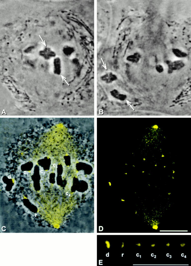
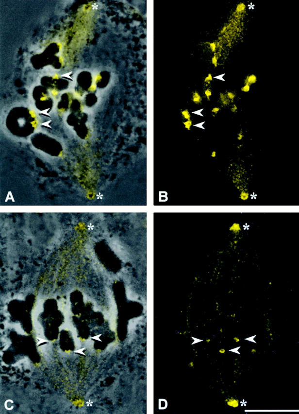
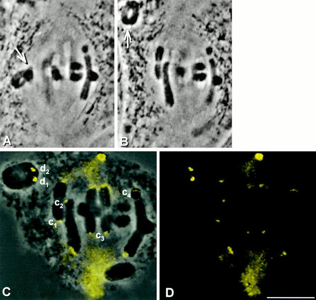
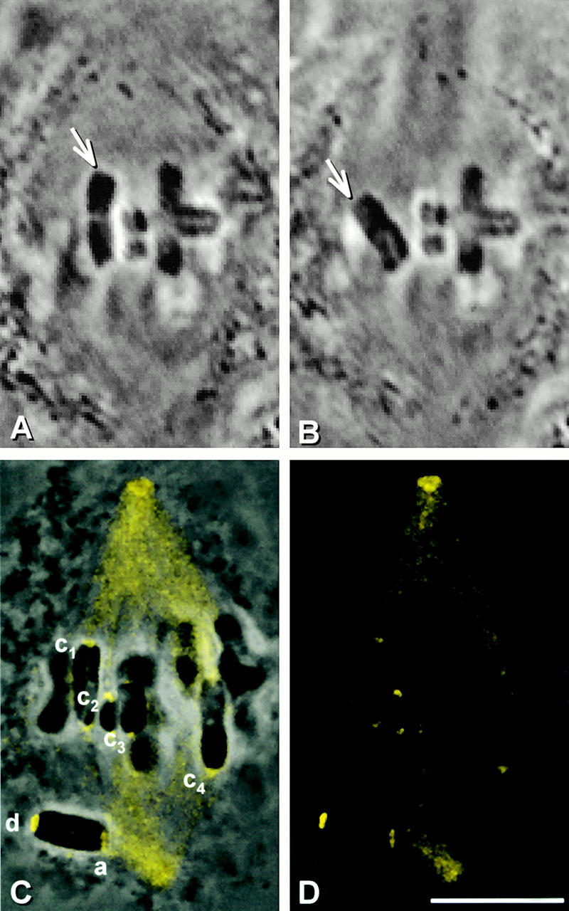
Cytoplasmic dynein is the only known kinetochore protein capable of driving chromosome movement toward spindle poles. In grasshopper spermatocytes, dynein immunofluorescence staining is bright at prometaphase kinetochores and dimmer at metaphase kinetochores. We have determined that these differences in staining intensity reflect differences in amounts of dynein associated with the kinetochore. Metaphase kinetochores regain bright dynein staining if they are detached from spindle microtubules by micromanipulation and kept detached for 10 min. We show that this increase in dynein staining is not caused by the retraction or unmasking of dynein upon detachment. Thus, dynein genuinely is a transient component of spermatocyte kinetochores. We further show that microtubule attachment, not tension, regulates dynein localization at kinetochores. Dynein binding is extremely sensitive to the presence of microtubules: fewer than half the normal number of kinetochore microtubules leads to the loss of most kinetochoric dynein. As a result, the bulk of the dynein leaves the kinetochore very early in mitosis, soon after the kinetochores begin to attach to microtubules. The possible functions of this dynein fraction are therefore limited to the initial attachment and movement of chromosomes and/or to a role in the mitotic checkpoint.
胞质动力蛋白是唯一已知的能够驱动染色体向纺锤体极移动的动粒蛋白。在蚱蜢精母细胞中,动力蛋白免疫荧光染色在前中期动粒处明亮,在中期动粒处较暗。我们已经确定,这些染色强度的差异反映了与动粒相关的动力蛋白数量的差异。如果通过显微操作将中期动粒从纺锤体微管上分离并保持分离10分钟,中期动粒会重新获得明亮的动力蛋白染色。我们表明,动力蛋白染色的这种增加不是由分离时动力蛋白的回缩或暴露引起的。因此,动力蛋白确实是精母细胞动粒的一个短暂成分。我们进一步表明,微管附着而非张力调节动力蛋白在动粒处的定位。动力蛋白的结合对微管的存在极为敏感:少于正常数量一半的动粒微管会导致大多数动粒动力蛋白的丢失。因此,大部分动力蛋白在有丝分裂早期,即动粒开始附着到微管后不久,就会离开动粒。因此,这种动力蛋白部分的可能功能仅限于染色体的初始附着和移动和/或在有丝分裂检查点中的作用。