• 文献检索
  • 文档翻译
  • 深度研究
  • 学术资讯
  • Suppr Zotero 插件Zotero 插件
  • 邀请有礼
  • 套餐&价格
  • 历史记录
应用&插件
Suppr Zotero 插件Zotero 插件浏览器插件Mac 客户端Windows 客户端微信小程序
定价
高级版会员购买积分包购买API积分包
服务
文献检索文档翻译深度研究API 文档MCP 服务
关于我们
关于 Suppr公司介绍联系我们用户协议隐私条款
关注我们

Suppr 超能文献

核心技术专利:CN118964589B侵权必究
粤ICP备2023148730 号-1Suppr @ 2026

文献检索

告别复杂PubMed语法,用中文像聊天一样搜索,搜遍4000万医学文献。AI智能推荐,让科研检索更轻松。

立即免费搜索

文件翻译

保留排版,准确专业,支持PDF/Word/PPT等文件格式,支持 12+语言互译。

免费翻译文档

深度研究

AI帮你快速写综述,25分钟生成高质量综述,智能提取关键信息,辅助科研写作。

立即免费体验

CT静脉成像在良性胸部中心静脉阻塞诊断与治疗中的作用

Role of CT venography in the diagnosis and treatment of benign thoracic central venous obstruction.

作者信息

Kim Hyo- Cheol, Chung Jin Wook, Park Jae Hyung, Yin Yong Hu, Park Seong Ho, Yoon Chang Jin, Choi Young Ho

机构信息

Department of Radiology, Seoul National University College of Medicine, Seoul, Korea.

出版信息

Korean J Radiol. 2003 Jul-Sep;4(3):146-52. doi: 10.3348/kjr.2003.4.3.146.

DOI:10.3348/kjr.2003.4.3.146
PMID:14530642
原文链接:https://pmc.ncbi.nlm.nih.gov/articles/PMC2698080/
Abstract

OBJECTIVE

To evaluate the role of CT venography in the diagnosis and treatment of benign thoracic central venous obstruction.

MATERIALS AND METHODS

Eighteen patients who had undergone both CT venography and digital subtraction venography were prospectively enrolled in this study. The following features were analyzed by two observers: the cause, degree, and extent of venous obstruction; associated thrombosis; and implications for the planning of treatment. CT venography and digital subtraction venography were compared in defined venous segments, and the degree of obstruction, and correlation was expressed using Spearman's rank correlation coefficient.

RESULTS

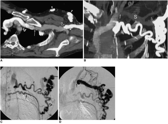
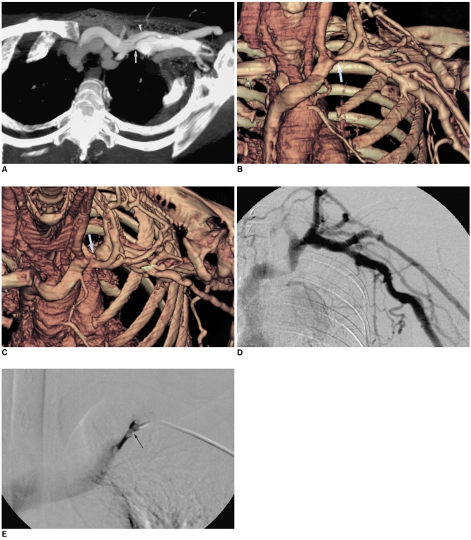
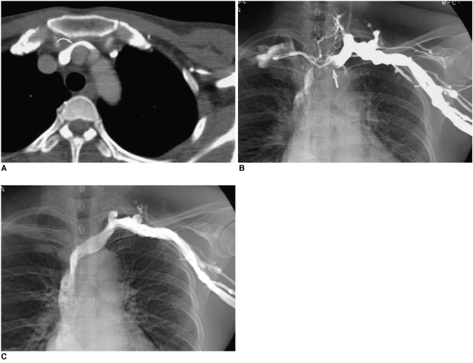
In all patients, CT venography depicted the causes of obstruction, including extrinsic compression of the left brachiocephalic vein, and mediastinal inflammatory pseudotumor. Interobserver agreement regarding classification of the degree of obstruction was judged as good for CT venography (K=0.864), and in evaluating this, there was significant correlation between CT venography and digital subtraction venography (reader 1: Rs = 0.58, p < 0.01; reader 2: Rs = 0.56, p < 0.01). In evaluating the status of central veins proximal to long segmental obstruction, and associated thrombosis, CT venography was superior to digital subtraction venography. In half of all patients, the findings of CT venography led to changes in the treatment plan.

CONCLUSION

The findings of CT venography correlated closely with those of digital subtraction venography, and the former accurately depicted the degree and extent of benign venous obstruction.

摘要

目的

评估CT静脉成像在良性胸段中心静脉梗阻诊断及治疗中的作用。

材料与方法

本前瞻性研究纳入了18例同时接受CT静脉成像和数字减影静脉造影的患者。两名观察者分析了以下特征:静脉梗阻的原因、程度及范围;相关血栓形成情况;以及对治疗方案规划的影响。在特定静脉节段对CT静脉成像和数字减影静脉造影进行比较,并使用Spearman等级相关系数表示梗阻程度及相关性。

结果

在所有患者中,CT静脉成像均显示了梗阻原因,包括左头臂静脉的外部压迫以及纵隔炎性假瘤。观察者间对CT静脉成像梗阻程度分类的一致性被判定为良好(K = 0.864),在评估这一点时,CT静脉成像与数字减影静脉造影之间存在显著相关性(观察者1:Rs = 0.58,p < 0.01;观察者2:Rs = 0.56,p < 0.01)。在评估长节段梗阻近端中心静脉的状况及相关血栓形成时,CT静脉成像优于数字减影静脉造影。在所有患者中,有一半患者CT静脉成像的结果导致了治疗方案的改变。

结论

CT静脉成像的结果与数字减影静脉造影的结果密切相关,前者能准确描绘良性静脉梗阻的程度及范围。

https://cdn.ncbi.nlm.nih.gov/pmc/blobs/72b4/2698080/3f83296ff707/kjr-4-146-g003.jpg
https://cdn.ncbi.nlm.nih.gov/pmc/blobs/72b4/2698080/dde15fd2d98b/kjr-4-146-g001.jpg
https://cdn.ncbi.nlm.nih.gov/pmc/blobs/72b4/2698080/d6428d262a78/kjr-4-146-g002.jpg
https://cdn.ncbi.nlm.nih.gov/pmc/blobs/72b4/2698080/3f83296ff707/kjr-4-146-g003.jpg
https://cdn.ncbi.nlm.nih.gov/pmc/blobs/72b4/2698080/dde15fd2d98b/kjr-4-146-g001.jpg
https://cdn.ncbi.nlm.nih.gov/pmc/blobs/72b4/2698080/d6428d262a78/kjr-4-146-g002.jpg
https://cdn.ncbi.nlm.nih.gov/pmc/blobs/72b4/2698080/3f83296ff707/kjr-4-146-g003.jpg

相似文献

1
Role of CT venography in the diagnosis and treatment of benign thoracic central venous obstruction.CT静脉成像在良性胸部中心静脉阻塞诊断与治疗中的作用
Korean J Radiol. 2003 Jul-Sep;4(3):146-52. doi: 10.3348/kjr.2003.4.3.146.
2
A three-dimensional gadolinium-enhanced MR venography technique for imaging central veins.一种用于中枢静脉成像的三维钆增强磁共振静脉造影技术。
AJR Am J Roentgenol. 1999 Oct;173(4):999-1003. doi: 10.2214/ajr.173.4.10511166.
3
Deep venous thrombosis with suspected pulmonary embolism: detection with combined CT venography and pulmonary angiography.疑似肺栓塞的深静脉血栓形成:联合CT静脉造影和肺血管造影检测
Radiology. 2001 May;219(2):498-502. doi: 10.1148/radiology.219.2.r01ma26498.
4
Complications of Permanent Hemodialysis Catheter Placement; Need for Better Pre-Implantation Algorithm?永久性血液透析导管置入的并发症;是否需要更好的植入前算法?
Ther Apher Dial. 2016 Aug;20(4):394-9. doi: 10.1111/1744-9987.12397. Epub 2016 Mar 28.
5
Membranous obstruction of the inferior vena cava: comparison of findings at sonography, CT, and venography.下腔静脉膜性梗阻:超声、CT及静脉造影检查结果比较
AJR Am J Roentgenol. 1992 Sep;159(3):515-20. doi: 10.2214/ajr.159.3.1503015.
6
Thoracic central venous evaluation: comparison of first-pass direct versus delayed-phase indirect multidetector CT venography.胸部中央静脉评估:直接第一通过法与间接多探测器 CT 静脉造影延迟期法的比较。
Clin Imaging. 2015 May-Jun;39(3):412-6. doi: 10.1016/j.clinimag.2015.02.005. Epub 2015 Feb 12.
7
Deep venous thrombosis: diagnosis by using venous enhanced subtracted peak arterial MR venography versus conventional venography.深静脉血栓形成:采用静脉增强减影峰值动脉磁共振静脉造影与传统静脉造影的诊断比较
Radiology. 2003 Mar;226(3):812-20. doi: 10.1148/radiol.2263012205. Epub 2003 Jan 24.
8
Thromboembolic disease: variability of interobserver agreement in the interpretation of CT venography with CT pulmonary angiography.
AJR Am J Roentgenol. 2001 Apr;176(4):1043-7. doi: 10.2214/ajr.176.4.1761043.
9
Evaluation of the lower extremity veins in patients with suspected pulmonary embolism: a retrospective comparison of helical CT venography and sonography. 2000 ARRS Executive Council Award I. American Roentgen Ray Society.疑似肺栓塞患者下肢静脉的评估:螺旋CT静脉造影与超声检查的回顾性比较。2000年ARRS执行委员会奖I。美国放射学会。
AJR Am J Roentgenol. 2000 Dec;175(6):1525-31. doi: 10.2214/ajr.175.6.1751525.
10
[The significance of magnetic resonance venography in the pre-interventional clarification of a malignant superior venous obstruction].
Rofo. 1998 Sep;169(3):253-9. doi: 10.1055/s-2007-1015086.

引用本文的文献

1
A Comprehensive Review of Catheter-Related Thrombosis.导管相关血栓形成的综合综述
J Clin Med. 2024 Dec 21;13(24):7818. doi: 10.3390/jcm13247818.
2
Interventional Therapy for Upper Extremity Phlegmasia Cerulea Dolens in a Patient With Heparin-Induced Thrombocytopenia: A Case Report.
Cureus. 2024 Apr 10;16(4):e57987. doi: 10.7759/cureus.57987. eCollection 2024 Apr.
3
Central venous stenosis after subclavian versus internal jugular dialysis catheter insertion (CITES) in adults in need of a temporary central dialysis catheter: study protocol for a two-arm, parallel-group, non-inferiority randomised controlled trial.成人临时中央透析导管需求患者锁骨下与颈内静脉置管后中心静脉狭窄(CITES):一项双臂、平行组、非劣效性随机对照试验的研究方案。

本文引用的文献

1
Superior vena cava stenting.上腔静脉支架置入术。
Radiol Clin North Am. 2000 Mar;38(2):409-24. doi: 10.1016/s0033-8389(05)70171-8.
2
Treatment of hemodialysis-related central venous stenosis or occlusion: results of primary Wallstent placement and follow-up in 50 patients.血液透析相关中心静脉狭窄或闭塞的治疗:50例患者初次置入Wallstent支架及随访结果
Radiology. 1999 Jul;212(1):175-80. doi: 10.1148/radiology.212.1.r99jl21175.
3
Helical CT phlebography of the superior vena cava: diagnosis and evaluation of venous obstruction.
Trials. 2023 May 12;24(1):327. doi: 10.1186/s13063-023-07350-9.
4
Pediatric Cardiothoracic CT Guideline Provided by the Asian Society of Cardiovascular Imaging Congenital Heart Disease Study Group: Part 2. Contemporary Clinical Applications.亚洲心血管影像学会先天性心脏病研究组制定的儿科心胸 CT 指南:第 2 部分。当代临床应用。
Korean J Radiol. 2021 Aug;22(8):1397-1415. doi: 10.3348/kjr.2020.1332. Epub 2021 May 4.
5
Diagnostic and Therapeutic Management of Upper Extremity Deep Vein Thrombosis.上肢深静脉血栓形成的诊断与治疗管理
J Clin Med. 2020 Jul 1;9(7):2069. doi: 10.3390/jcm9072069.
6
Computed tomography venography of the upper extremities - Using low dose bilateral contrast media injection in a patient with suspected venous thoracic outlet syndrome.上肢计算机断层扫描静脉造影——对疑似胸廓出口静脉综合征患者使用低剂量双侧造影剂注射
Radiol Case Rep. 2020 Jan 15;15(3):302-305. doi: 10.1016/j.radcr.2019.12.023. eCollection 2020 Mar.
7
Venous Obstruction in Cardiac Rhythm Device Therapy.心脏节律装置治疗中的静脉阻塞
Curr Treat Options Cardiovasc Med. 2018 Jul 11;20(8):64. doi: 10.1007/s11936-018-0664-5.
8
Comparison of Covered Versus Uncovered Stents for Benign Superior Vena Cava (SVC) Obstruction.覆膜支架与非覆膜支架治疗良性上腔静脉阻塞的比较。
Cardiovasc Intervent Radiol. 2018 May;41(5):712-717. doi: 10.1007/s00270-018-1906-3. Epub 2018 Feb 28.
9
Diagnostic value of computerized tomography venography in detecting stenosis and occlusion of subclavian vein and superior vena in chronic renal failure patients.计算机断层扫描静脉成像在检测慢性肾衰竭患者锁骨下静脉和上腔静脉狭窄及闭塞中的诊断价值
Electron Physician. 2016 Aug 25;8(8):2781-2786. doi: 10.19082/2781. eCollection 2016 Aug.
10
MDCT Venography Evaluation of a Rare Collateral Vein Draining from the Left Subclavian Vein to the Great Cardiac Vein.多层螺旋CT静脉成像评估一条从左锁骨下静脉引流至心大静脉的罕见侧支静脉
J Clin Imaging Sci. 2014 Oct 27;4:58. doi: 10.4103/2156-7514.143425. eCollection 2014.
上腔静脉螺旋CT静脉造影:静脉阻塞的诊断与评估
AJR Am J Roentgenol. 1999 May;172(5):1327-33. doi: 10.2214/ajr.172.5.10227511.
4
Use of Wallstents for hemodialysis access-related venous stenoses and occlusions untreatable with balloon angioplasty.使用Wallstents治疗经球囊血管成形术无法治疗的血液透析通路相关静脉狭窄和闭塞。
Radiology. 1995 May;195(2):479-84. doi: 10.1148/radiology.195.2.7724770.
5
Etiologic considerations in superior vena cava syndrome.上腔静脉综合征的病因学考量
Mayo Clin Proc. 1981 Jul;56(7):407-13.
6
Hemodialysis-associated subclavian vein stenosis.血液透析相关的锁骨下静脉狭窄
Kidney Int. 1988 Jun;33(6):1156-9. doi: 10.1038/ki.1988.124.
7
Axillary and subclavian vein stenosis: percutaneous angioplasty.
Radiology. 1988 Aug;168(2):371-3. doi: 10.1148/radiology.168.2.2969117.
8
The role of percutaneous angioplasty in the management of chronic hemodialysis fistulas.
Ann Surg. 1987 Dec;206(6):777-81. doi: 10.1097/00000658-198712000-00015.
9
The importance of preoperative evaluation of the subclavian vein in dialysis access planning.锁骨下静脉术前评估在透析通路规划中的重要性。
AJR Am J Roentgenol. 1991 Mar;156(3):623-5. doi: 10.2214/ajr.156.3.1781814.
10
Compression of the left brachiocephalic vein by the innominate artery resulting in massive arm edema in a hemodialysis patient.
Am J Nephrol. 1992;12(1-2):108-10. doi: 10.1159/000168427.