Mann Christopher J, Yu Lingfeng, Kim Myung K
Department of Physics, University of South Florida, Tampa, FL 33620, USA.
Biomed Eng Online. 2006 Mar 23;5:21. doi: 10.1186/1475-925X-5-21.
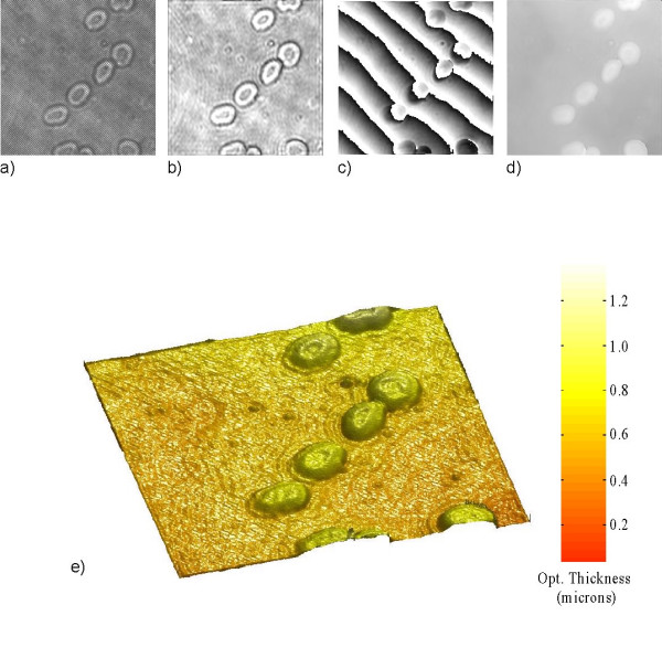
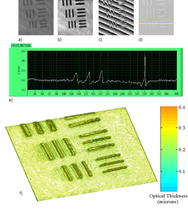
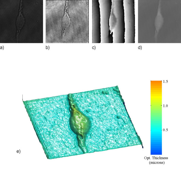
Many biological specimens, such as living cells and their intracellular components, often exhibit very little amplitude contrast, making it difficult for conventional bright field microscopes to distinguish them from their surroundings. To overcome this problem phase contrast techniques such as Zernike, Normarsky and dark-field microscopies have been developed to improve specimen visibility without chemically or physically altering them by the process of staining. These techniques have proven to be invaluable tools for studying living cells and furthering scientific understanding of fundamental cellular processes such as mitosis. However a drawback of these techniques is that direct quantitative phase imaging is not possible. Quantitative phase imaging is important because it enables determination of either the refractive index or optical thickness variations from the measured optical path length with sub-wavelength accuracy. Digital holography is an emergent phase contrast technique that offers an excellent approach in obtaining both qualitative and quantitative phase information from the hologram. A CCD camera is used to record a hologram onto a computer and numerical methods are subsequently applied to reconstruct the hologram to enable direct access to both phase and amplitude information. Another attractive feature of digital holography is the ability to focus on multiple focal planes from a single hologram, emulating the focusing control of a conventional microscope.
A modified Mach-Zender off-axis setup in transmission is used to record and reconstruct a number of holographic amplitude and phase images of cellular and sub-cellular features.
Both cellular and sub-cellular features are imaged with sub-micron, diffraction-limited resolution. Movies of holographic amplitude and phase images of living microbes and cells are created from a series of holograms and reconstructed with numerically adjustable focus, so that the moving object can be accurately tracked with a reconstruction rate of 300ms for each hologram. The holographic movies show paramecium swimming among other microbes as well as displaying some of their intracellular processes. A time lapse movie is also shown for fibroblast cells in the process of migration.
Digital holography and movies of digital holography are seen to be useful new tools for visualization of dynamic processes in biological microscopy. Phase imaging digital holography is a promising technique in terms of the lack of coherent noise and the precision with which the optical thickness of a sample can be profiled, which can lead to images with an axial resolution of a few nanometres.
许多生物标本,如活细胞及其细胞内成分,通常表现出非常小的振幅对比度,这使得传统的明场显微镜很难将它们与周围环境区分开来。为了克服这个问题,已经开发了诸如泽尼克相衬、诺马斯基相衬和暗场显微镜等相衬技术,以提高标本的可见性,而无需通过染色过程对其进行化学或物理改变。这些技术已被证明是研究活细胞和深化对有丝分裂等基本细胞过程的科学理解的宝贵工具。然而,这些技术的一个缺点是无法进行直接的定量相成像。定量相成像很重要,因为它能够以亚波长精度根据测量的光程长度确定折射率或光学厚度的变化。数字全息术是一种新兴的相衬技术,它为从全息图中获取定性和定量相信息提供了一种出色的方法。使用电荷耦合器件(CCD)相机将全息图记录到计算机上,随后应用数值方法重建全息图,以便直接获取相位和振幅信息。数字全息术的另一个吸引人的特点是能够从单个全息图聚焦到多个焦平面,模拟传统显微镜的聚焦控制。
使用一种改进的马赫 - 曾德尔离轴透射设置来记录和重建细胞及亚细胞特征的多个全息振幅和相位图像。
细胞和亚细胞特征均以亚微米级的衍射极限分辨率成像。从一系列全息图创建活微生物和细胞的全息振幅和相位图像的影片,并通过数值可调聚焦进行重建,这样对于每个全息图,以300毫秒的重建速率可以准确跟踪移动物体。全息影片展示了草履虫在其他微生物中游动以及一些细胞内过程。还展示了成纤维细胞迁移过程的延时影片。
数字全息术及数字全息影片被视为生物显微镜中可视化动态过程的有用新工具。相成像数字全息术是一种很有前景的技术,因为它缺乏相干噪声,并且能够精确测量样品的光学厚度,这可以产生轴向分辨率为几纳米的图像。