• 文献检索
  • 文档翻译
  • 深度研究
  • 学术资讯
  • Suppr Zotero 插件Zotero 插件
  • 邀请有礼
  • 套餐&价格
  • 历史记录
应用&插件
Suppr Zotero 插件Zotero 插件浏览器插件Mac 客户端Windows 客户端微信小程序
定价
高级版会员购买积分包购买API积分包
服务
文献检索文档翻译深度研究API 文档MCP 服务
关于我们
关于 Suppr公司介绍联系我们用户协议隐私条款
关注我们

Suppr 超能文献

核心技术专利:CN118964589B侵权必究
粤ICP备2023148730 号-1Suppr @ 2026

文献检索

告别复杂PubMed语法,用中文像聊天一样搜索,搜遍4000万医学文献。AI智能推荐,让科研检索更轻松。

立即免费搜索

文件翻译

保留排版,准确专业,支持PDF/Word/PPT等文件格式,支持 12+语言互译。

免费翻译文档

深度研究

AI帮你快速写综述,25分钟生成高质量综述,智能提取关键信息,辅助科研写作。

立即免费体验

坦桑尼亚CT检查中患者器官剂量的估算。

Estimation of patient organ doses from CT examinations in Tanzania.

作者信息

Ngaile Justin E, Msaki Peter K

机构信息

Radiation Control Directorate, Tanzania Atomic Energy Commission, Arusha, Tanzania.

出版信息

J Appl Clin Med Phys. 2006 Aug 24;7(3):80-94. doi: 10.1120/jacmp.v7i3.2200.

DOI:10.1120/jacmp.v7i3.2200
PMID:17533338
原文链接:https://pmc.ncbi.nlm.nih.gov/articles/PMC5722432/
Abstract

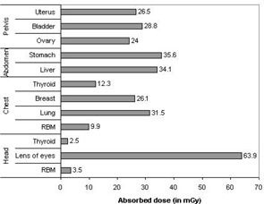
Although the use of computed tomography (CT) in medical diagnosis delivers relatively higher radiation doses to patients than other radiological procedures, lack of optimized protocols could be an additional source of increased dose in developing countries. The aim of this study was to determine the magnitude of radiation doses received by selected radiosensitive organs of patients from CT examinations. The study was further carried out in order to assess the influence of existing CT scanning protocols on patient organ doses. In order to achieve these objectives, patient organ doses from five common CT examinations were obtained from eight hospitals in Tanzania. The patient organ doses were estimated using measurements of CTDI, exposure-related parameters and the NRPB conversion factors. Large variation of mean organ doses among hospitals was observed for similar CT examinations. These variations were largely originated from different CT scanning protocols employed in different hospitals and scanner type. The mean organ doses in this study for the lens of the eyes (for head), thyroid (for chest), breast (for chest), stomach (for abdomen), and ovary (for pelvis), were 63.9 mGy, 12.3 mGy, 26.1 mGy, 35.6 mGy, and 24.0 mGy, respectively. These values were mostly comparable and slightly higher than the values of organ doses reported from literature for the UK, Japan, Germany, Norway and the Netherlands. It was concluded that patient organ doses could substantially minimized through careful selection of scanning parameters based on clinical indications of study, patient size, and body region being examined. Additional dose reduction to superficial organs would require the use of shielding materials.

摘要

尽管在医学诊断中使用计算机断层扫描(CT)给患者带来的辐射剂量比其他放射学检查相对更高,但在发展中国家,缺乏优化的方案可能是剂量增加的另一个来源。本研究的目的是确定接受CT检查的患者选定的辐射敏感器官所接受的辐射剂量大小。进一步开展该研究是为了评估现有CT扫描方案对患者器官剂量的影响。为了实现这些目标,从坦桑尼亚的八家医院获取了五种常见CT检查的患者器官剂量。使用CTDI测量值、与曝光相关的参数和NRPB转换因子来估计患者器官剂量。对于类似的CT检查,观察到不同医院之间平均器官剂量存在很大差异。这些差异主要源于不同医院采用的不同CT扫描方案和扫描仪类型。本研究中,眼睛晶状体(头部检查)、甲状腺(胸部检查)、乳房(胸部检查)、胃(腹部检查)和卵巢(骨盆检查)的平均器官剂量分别为63.9毫戈瑞、12.3毫戈瑞、26.1毫戈瑞、35.6毫戈瑞和24.0毫戈瑞。这些值大多具有可比性,且略高于英国、日本、德国、挪威和荷兰文献报道的器官剂量值。得出的结论是,通过根据研究的临床指征、患者体型和被检查的身体部位仔细选择扫描参数,可以大幅降低患者器官剂量。对浅表器官进一步降低剂量将需要使用屏蔽材料。

https://cdn.ncbi.nlm.nih.gov/pmc/blobs/53d6/5722432/0ad119345de3/ACM2-7-80-g006.jpg
https://cdn.ncbi.nlm.nih.gov/pmc/blobs/53d6/5722432/66705563fd4e/ACM2-7-80-g001.jpg
https://cdn.ncbi.nlm.nih.gov/pmc/blobs/53d6/5722432/ecdbc83b2dbe/ACM2-7-80-g002.jpg
https://cdn.ncbi.nlm.nih.gov/pmc/blobs/53d6/5722432/d957de4bdabb/ACM2-7-80-g003.jpg
https://cdn.ncbi.nlm.nih.gov/pmc/blobs/53d6/5722432/7193b76dd4f0/ACM2-7-80-g004.jpg
https://cdn.ncbi.nlm.nih.gov/pmc/blobs/53d6/5722432/1123a18a01f4/ACM2-7-80-g005.jpg
https://cdn.ncbi.nlm.nih.gov/pmc/blobs/53d6/5722432/0ad119345de3/ACM2-7-80-g006.jpg

相似文献

1
Estimation of patient organ doses from CT examinations in Tanzania.坦桑尼亚CT检查中患者器官剂量的估算。
J Appl Clin Med Phys. 2006 Aug 24;7(3):80-94. doi: 10.1120/jacmp.v7i3.2200.
2
Current status of patient radiation doses from computed tomography examinations in Tanzania.坦桑尼亚计算机断层扫描检查中患者辐射剂量的现状
Radiat Prot Dosimetry. 2006;121(2):128-35. doi: 10.1093/rpd/ncl001. Epub 2006 Feb 3.
3
Towards establishment of the national reference dose levels from computed tomography examinations in Tanzania.迈向确定坦桑尼亚计算机断层扫描检查的国家参考剂量水平。
J Radiol Prot. 2006 Jun;26(2):213-25. doi: 10.1088/0952-4746/26/2/006. Epub 2006 May 26.
4
Patient size and radiation exposure in thoracic, pelvic, and abdominal CT examinations performed with automatic exposure control.自动曝光控制在胸部、盆腔和腹部 CT 检查中对患者体型和辐射暴露的影响。
AJR Am J Roentgenol. 2010 Dec;195(6):1342-6. doi: 10.2214/AJR.09.3331.
5
Toward automated and personalized organ dose determination in CT examinations - A comparison of two tissue characterization models for Monte Carlo organ dose calculation with a Therapy Planning System.迈向 CT 检查中自动化和个性化器官剂量测定——两种组织特征化模型在治疗计划系统中进行蒙特卡罗器官剂量计算的比较。
Med Phys. 2019 Feb;46(2):1012-1023. doi: 10.1002/mp.13357. Epub 2019 Jan 15.
6
Organ doses for reference pediatric and adolescent patients undergoing computed tomography estimated by Monte Carlo simulation.基于蒙特卡罗模拟计算的参考小儿和青少年患者 CT 检查器官剂量。
Med Phys. 2012 Apr;39(4):2129-46. doi: 10.1118/1.3693052.
7
Radiation doses to patients during selected CT procedures at four hospitals in Tanzania.坦桑尼亚四家医院在选定的CT检查过程中患者所接受的辐射剂量。
Eur J Radiol. 2006 Mar;57(3):461-7. doi: 10.1016/j.ejrad.2005.08.003. Epub 2005 Sep 13.
8
Major trauma & cervical clearance radiation doses & cancer induction.严重创伤、颈椎清除放疗剂量与癌症诱发
Injury. 2008 Mar;39(3):347-56. doi: 10.1016/j.injury.2007.06.013. Epub 2007 Oct 4.
9
Organ doses evaluation for chest computed tomography procedures with TL dosimeters: Comparison with Monte Carlo simulations.使用热释光剂量计对胸部计算机断层扫描程序进行器官剂量评估:与蒙特卡罗模拟的比较。
J Appl Clin Med Phys. 2019 Jan;20(1):308-320. doi: 10.1002/acm2.12505. Epub 2018 Dec 3.
10
Conversion factors to derive organ doses for canine subjects undergoing CT examinations.用于推导接受 CT 检查的犬科动物器官剂量的转换因子。
Vet Radiol Ultrasound. 2021 Jul;62(4):421-428. doi: 10.1111/vru.12979. Epub 2021 May 14.

引用本文的文献

1
Representative Organ Doses from Computed Tomography (CT) Exams from a Large International Registry.来自大型国际登记处的计算机断层扫描(CT)检查的代表性器官剂量。
Radiat Res. 2025 Jan 1;203(1):1-9. doi: 10.1667/RADE-24-00178.1.
2
Assessment and comparison of radiation dose and image quality in multi-detector CT scanners in non-contrast head and neck examinations.多排螺旋CT扫描仪在头颈部非增强检查中辐射剂量与图像质量的评估及比较
Pol J Radiol. 2019 Jan 23;84:e61-e67. doi: 10.5114/pjr.2019.82743. eCollection 2019.
3
A survey of organ equivalent and effective doses from diagnostic radiology procedures.

本文引用的文献

1
A method to obtain the same levels of CT image noise for patients of various sizes, to minimize radiation dose.一种为不同体型患者获取相同水平CT图像噪声以最小化辐射剂量的方法。
Br J Radiol. 2002 Feb;75(890):140-50. doi: 10.1259/bjr.75.890.750140.
2
Managing patient dose in computed tomography. A report of the International Commission on Radiological Protection.计算机断层扫描中的患者剂量管理。国际放射防护委员会报告。
Ann ICRP. 2000;30(4):7-45. doi: 10.1016/s0146-6453(01)00049-5.
3
The potential for reduction of radiation doses to patients undergoing some common X ray examinations in Tanzania.
一项关于诊断性放射学检查中器官等效剂量和有效剂量的调查。
ISRN Radiol. 2012 Sep 6;2013:204346. doi: 10.5402/2013/204346. eCollection 2013.
坦桑尼亚一些常见X射线检查中降低患者辐射剂量的可能性。
Radiat Prot Dosimetry. 2001;94(4):381-4. doi: 10.1093/oxfordjournals.rpd.a006514.
4
Radioprotection to the eye during CT scanning.CT扫描期间对眼睛的辐射防护。
AJNR Am J Neuroradiol. 2001 Jun-Jul;22(6):1194-8.
5
Minimizing radiation dose for pediatric body applications of single-detector helical CT: strategies at a large Children's Hospital.单探测器螺旋CT在儿科身体检查中的辐射剂量最小化:一家大型儿童医院的策略
AJR Am J Roentgenol. 2001 Feb;176(2):303-6. doi: 10.2214/ajr.176.2.1760303.
6
Estimated risks of radiation-induced fatal cancer from pediatric CT.儿童CT辐射诱发致命癌症的估计风险。
AJR Am J Roentgenol. 2001 Feb;176(2):289-96. doi: 10.2214/ajr.176.2.1760289.
7
Effective doses to patients undergoing thoracic computed tomography examinations.胸部计算机断层扫描检查患者的有效剂量。
Med Phys. 2000 May;27(5):838-44. doi: 10.1118/1.598949.
8
Relationships between physical dose quantities and patient dose in CT.CT中物理剂量量与患者剂量之间的关系。
Br J Radiol. 1999 Jun;72(858):556-61. doi: 10.1259/bjr.72.858.10560337.
9
Dosimetry for optimisation of patient protection in computed tomography.计算机断层扫描中患者防护优化的剂量测定法。
Appl Radiat Isot. 1999 Jan;50(1):165-72. doi: 10.1016/s0969-8043(98)00024-4.
10
The breast: in-plane x-ray protection during diagnostic thoracic CT--shielding with bismuth radioprotective garments.乳房:诊断性胸部CT扫描期间的平面X线防护——使用铋辐射防护衣进行屏蔽
Radiology. 1997 Dec;205(3):853-8. doi: 10.1148/radiology.205.3.9393547.