Seko Yuko, Azuma Noriyuki, Takahashi Yoriko, Makino Hatsune, Morito Toshiyuki, Muneta Takeshi, Matsumoto Kenji, Saito Hirohisa, Sekiya Ichiro, Umezawa Akihiro
Department of Reproductive Biology and Pathology, National Institute for Child and Health Development, Tokyo, Japan.
PLoS One. 2008;3(11):e3709. doi: 10.1371/journal.pone.0003709. Epub 2008 Nov 12.
The sclera maintains and protects the eye ball, which receives visual inputs. Although the sclera does not contribute significantly to visual perception, scleral diseases such as refractory scleritis, scleral perforation and pathological myopia are considered incurable or difficult to cure. The aim of this study is to identify characteristics of the human sclera as one of the connective tissues derived from the neural crest and mesoderm.
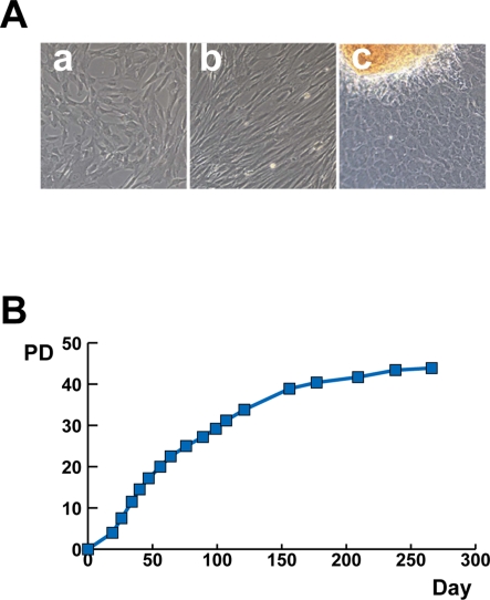
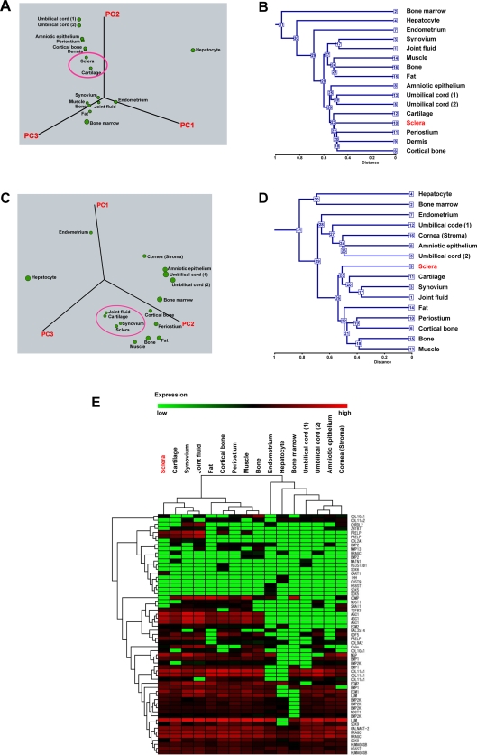
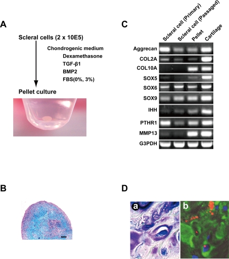
METHODOLOGY/PRINCIPAL FINDINGS: We have demonstrated microarray data of cultured human infant scleral cells. Hierarchical clustering was performed to group scleral cells and other mesenchymal cells into subcategories. Hierarchical clustering analysis showed similarity between scleral cells and auricular cartilage-derived cells. Cultured micromasses of scleral cells exposed to TGF-betas and BMP2 produced an abundant matrix. The expression of cartilage-associated genes, such as Indian hedge hog, type X collagen, and MMP13, was up-regulated within 3 weeks in vitro. These results suggest that human 'sclera'-derived cells can be considered chondrocytes when cultured ex vivo.
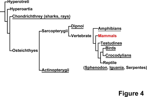
CONCLUSIONS/SIGNIFICANCE: Our present study shows a chondrogenic potential of human sclera. Interestingly, the sclera of certain vertebrates, such as birds and fish, is composed of hyaline cartilage. Although the human sclera is not a cartilaginous tissue, the human sclera maintains chondrogenic potential throughout evolution. In addition, our findings directly explain an enigma that the sclera and the joint cartilage are common targets of inflammatory cells in rheumatic arthritis. The present global gene expression database will contribute to the clarification of the pathogenesis of developmental diseases such as high myopia.
巩膜维持并保护眼球,眼球接收视觉输入。尽管巩膜对视觉感知的贡献不大,但诸如难治性巩膜炎、巩膜穿孔和病理性近视等巩膜疾病被认为无法治愈或难以治愈。本研究的目的是确定人类巩膜作为源自神经嵴和中胚层的结缔组织之一的特征。
方法/主要发现:我们展示了培养的人类婴儿巩膜细胞的微阵列数据。进行层次聚类以将巩膜细胞和其他间充质细胞分组为亚类。层次聚类分析显示巩膜细胞与耳软骨来源的细胞之间存在相似性。暴露于转化生长因子β和骨形态发生蛋白2的巩膜细胞培养微团产生了丰富的基质。软骨相关基因,如印度刺猬因子、X型胶原蛋白和基质金属蛋白酶13的表达在体外3周内上调。这些结果表明,人类“巩膜”来源的细胞在体外培养时可被视为软骨细胞。
结论/意义:我们目前的研究显示了人类巩膜的软骨形成潜力。有趣的是,某些脊椎动物,如鸟类和鱼类的巩膜由透明软骨组成。虽然人类巩膜不是软骨组织,但人类巩膜在整个进化过程中都保持着软骨形成潜力。此外,我们的发现直接解释了一个谜团,即巩膜和关节软骨是风湿性关节炎中炎症细胞的共同靶点。目前的全球基因表达数据库将有助于阐明诸如高度近视等高发性疾病的发病机制。