Center for In Vivo Microscopy, Duke University, Durham, North Carolina, USA.
Magn Reson Med. 2010 Apr;63(4):979-87. doi: 10.1002/mrm.22259.
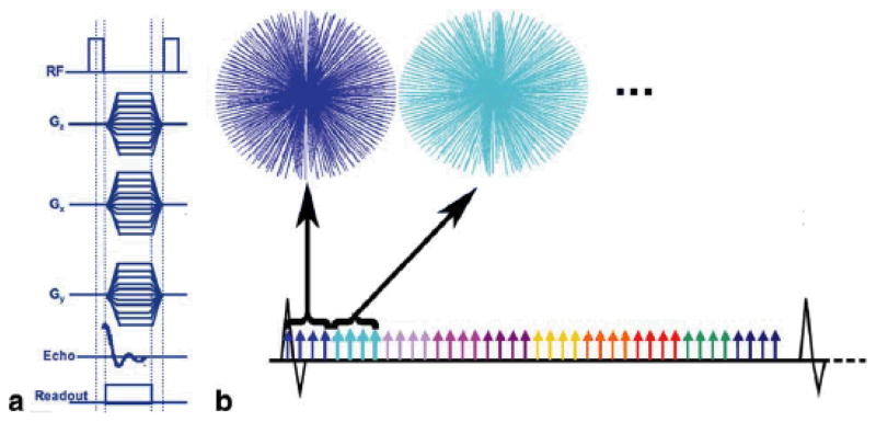
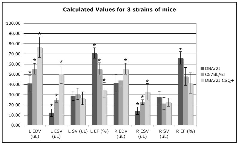
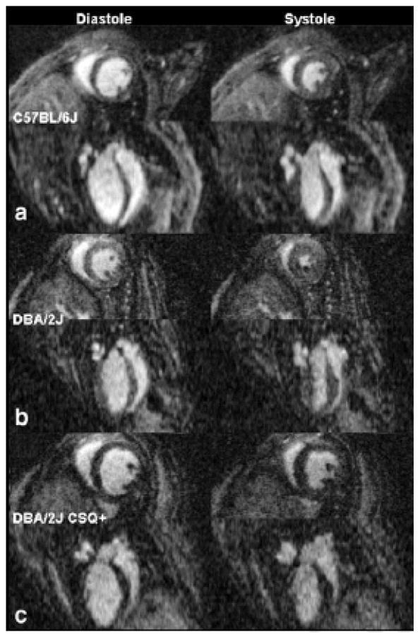
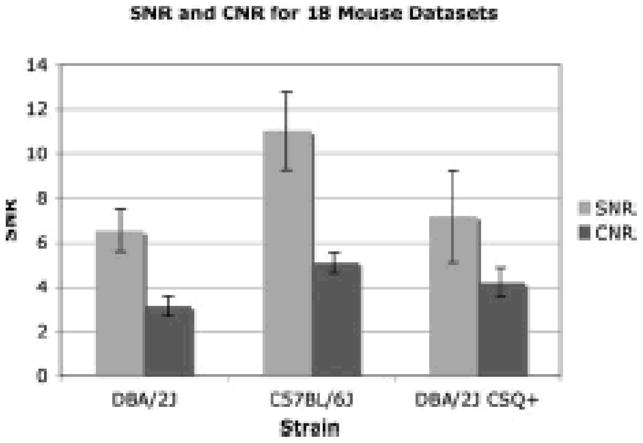
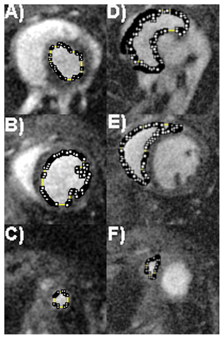
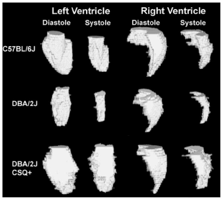
MR microscopy has enormous potential for small-animal cardiac imaging because it is capable of producing volumetric images at multiple time points to accurately measure cardiac function. MR has not been used as frequently as ultrasound to measure cardiac function in the small animal because the MR methods required relatively long scan times, limiting throughput. Here, we demonstrate four-dimensional radial acquisition in conjunction with a liposomal blood pool agent to explore functional differences in three populations of mice: six C57BL/6J mice, six DBA/2J mice, and six DBA/2J CSQ+ mice, all with the same gestational age and approximately the same weight. Cardiovascular function was determined by measuring both left ventricular and right ventricular end diastolic volume, end systolic volume, stroke volume, and ejection fraction. Statistical significance was observed in end diastolic volume, end systolic volume, and ejection fraction for left ventricular measurements between all three populations of mice. No statistically significant difference was observed in stroke volume in either the left or right ventricle for any of the three populations of mice. This study shows that MRI is capable of efficient, high-throughput, four-dimensional cardiovascular phenotyping of the mouse.
磁共振显微镜在小动物心脏成像方面具有巨大的潜力,因为它能够在多个时间点生成容积图像,从而准确测量心脏功能。磁共振成像(MRI)并没有像超声那样频繁地用于小动物心脏功能的测量,因为 MRI 方法需要相对较长的扫描时间,限制了吞吐量。在这里,我们展示了四维径向采集与脂质体血池造影剂相结合,以探索三种小鼠群体之间的功能差异:六只 C57BL/6J 小鼠、六只 DBA/2J 小鼠和六只 DBA/2J CSQ+ 小鼠,它们具有相同的胎龄和大致相同的体重。通过测量左心室和右心室舒张末期容积、收缩末期容积、每搏输出量和射血分数来确定心血管功能。在左心室测量中,三种小鼠群体之间在舒张末期容积、收缩末期容积和射血分数方面都观察到了统计学意义上的显著差异。在左心室或右心室的每搏输出量方面,三种小鼠群体之间均未观察到统计学上的显著差异。这项研究表明,MRI 能够高效、高通量地对小鼠进行四维心血管表型分析。