Rahman Mohammed S, Ingole Nilesh, Roblyer Darren, Stepanek Vanda, Richards-Kortum Rebecca, Gillenwater Ann, Shastri Surendra, Chaturvedi Pankaj
Department of Bioengineering, Rice University, Houston, 77005, USA.
Head and Neck Surgery, Tata Memorial Hospital, Mumbai, 400012, India.
Head Neck Oncol. 2010 Apr 22;2:10. doi: 10.1186/1758-3284-2-10.
There is an important global need to improve early detection of oral cancer. Recent reports suggest that optical imaging technologies can aid in the identification of neoplastic lesions in the oral cavity; however, there is little data evaluating the use of optical imaging modalities in resource limited settings where oral cancer impacts patients disproportionately. In this article, we evaluate a simple, low-cost optical imaging system that is designed for early detection of oral cancer in resource limited settings. We report results of a clinical study conducted at Tata Memorial Hospital (TMH) in Mumbai, India using this system as a tool to improve detection of oral cancer and its precursors.
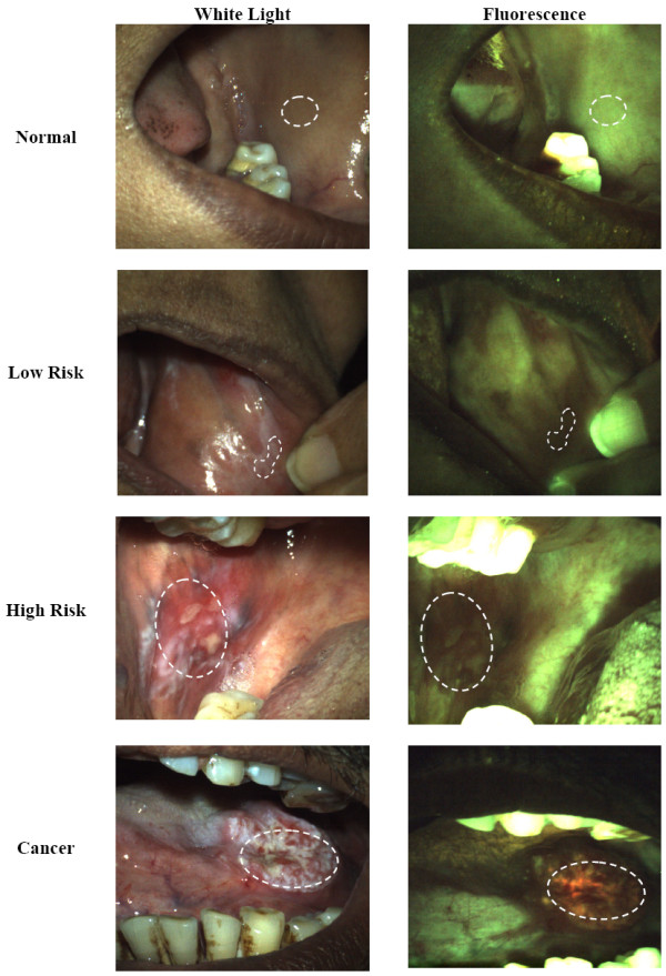
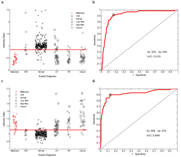
Reflectance images with white light illumination and fluorescence images with 455 nm excitation were obtained from 261 sites in the oral cavity from 76 patients and 90 sites in the oral cavity from 33 normal volunteers. Quantitative image features were used to develop classification algorithms to identify neoplastic tissue, using clinical diagnosis of expert observers as the gold standard.
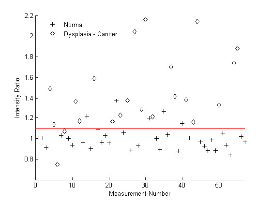
Using the ratio of red to green autofluorescence, the algorithm identified tissues judged clinically to be cancer or clinically suspicious for neoplasia with a sensitivity of 90% and a specificity of 87%.
Results suggest that the performance of this simple, objective low-cost system has potential to improve oral screening efforts, especially in low-resource settings.
全球迫切需要改进口腔癌的早期检测。最近的报告表明,光学成像技术有助于识别口腔中的肿瘤性病变;然而,在口腔癌对患者影响尤为严重的资源有限环境中,评估光学成像模式使用情况的数据很少。在本文中,我们评估了一种简单、低成本的光学成像系统,该系统旨在用于资源有限环境下口腔癌的早期检测。我们报告了在印度孟买塔塔纪念医院(TMH)进行的一项临床研究结果,该研究使用该系统作为改善口腔癌及其癌前病变检测的工具。
从76例患者口腔中的261个部位以及33名正常志愿者口腔中的90个部位获取白光照明反射图像和455nm激发荧光图像。以专家观察者的临床诊断为金标准,使用定量图像特征开发分类算法以识别肿瘤组织。
利用红色与绿色自发荧光的比率,该算法识别出临床判断为癌症或临床可疑为肿瘤的组织,灵敏度为90%,特异性为87%。
结果表明,这种简单、客观的低成本系统的性能有潜力改善口腔筛查工作,尤其是在资源匮乏的环境中。