• 文献检索
  • 文档翻译
  • 深度研究
  • 学术资讯
  • Suppr Zotero 插件Zotero 插件
  • 邀请有礼
  • 套餐&价格
  • 历史记录
应用&插件
Suppr Zotero 插件Zotero 插件浏览器插件Mac 客户端Windows 客户端微信小程序
定价
高级版会员购买积分包购买API积分包
服务
文献检索文档翻译深度研究API 文档MCP 服务
关于我们
关于 Suppr公司介绍联系我们用户协议隐私条款
关注我们

Suppr 超能文献

核心技术专利:CN118964589B侵权必究
粤ICP备2023148730 号-1Suppr @ 2026

文献检索

告别复杂PubMed语法,用中文像聊天一样搜索,搜遍4000万医学文献。AI智能推荐,让科研检索更轻松。

立即免费搜索

文件翻译

保留排版,准确专业,支持PDF/Word/PPT等文件格式,支持 12+语言互译。

免费翻译文档

深度研究

AI帮你快速写综述,25分钟生成高质量综述,智能提取关键信息,辅助科研写作。

立即免费体验

肾脏组织内的折射率不均匀性对多光子荧光激发显微镜的影响。

The effects of refractive index heterogeneity within kidney tissue on multiphoton fluorescence excitation microscopy.

机构信息

Department of Medicine, Division of Nephrology, Indiana University School of Medicine, Indianapolis, Indiana 46202–5188, USA.

出版信息

J Microsc. 2011 May;242(2):148-56. doi: 10.1111/j.1365-2818.2010.03448.x. Epub 2010 Sep 27.

DOI:10.1111/j.1365-2818.2010.03448.x
PMID:21118239
原文链接:https://pmc.ncbi.nlm.nih.gov/articles/PMC4450360/
Abstract

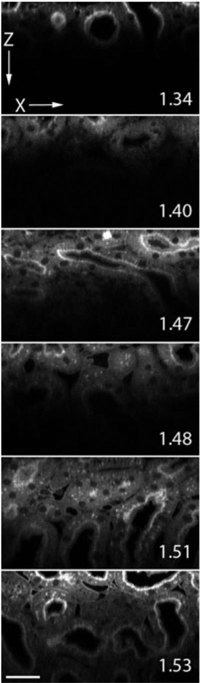
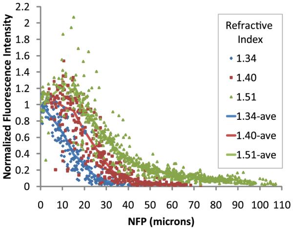
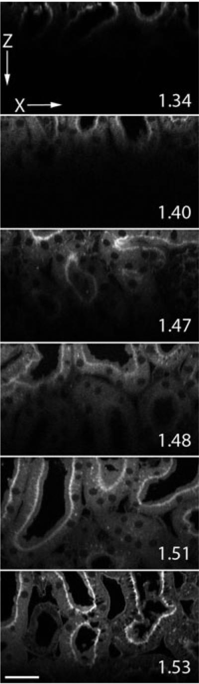
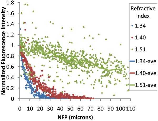
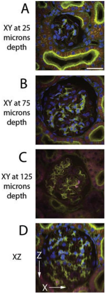
Although multiphoton fluorescence excitation microscopy has improved the depth at which useful fluorescence images can be collected in biological tissues, the reach of multiphoton fluorescence excitation microscopy is nonetheless limited by tissue scattering and spherical aberration. Scattering can be reduced in fixed samples by mounting in a medium whose refractive index closely matches that of the fixed material. Using optical 'clearing', the effects of refractive index heterogeneity on signal attenuation with depth are investigated. Quantitative measurements show that by mounting kidney tissue in a high refractive index medium, less than 50% of signal attenuates in 100 μm of depth.

摘要

尽管多光子荧光激发显微镜提高了在生物组织中收集有用荧光图像的深度,但多光子荧光激发显微镜的探测深度仍然受到组织散射和球差的限制。在固定样本中,可以通过在折射率与固定材料非常接近的介质中进行安装来减少散射。使用光学“透明化”技术,研究了折射率异质性对信号随深度衰减的影响。定量测量表明,通过将肾脏组织安装在高折射率介质中,在 100μm 的深度内,信号衰减不到 50%。

https://cdn.ncbi.nlm.nih.gov/pmc/blobs/343d/4450360/86a0a9976c17/nihms-693490-f0007.jpg
https://cdn.ncbi.nlm.nih.gov/pmc/blobs/343d/4450360/6fffd6eacdf2/nihms-693490-f0001.jpg
https://cdn.ncbi.nlm.nih.gov/pmc/blobs/343d/4450360/bb0843d136a0/nihms-693490-f0002.jpg
https://cdn.ncbi.nlm.nih.gov/pmc/blobs/343d/4450360/0d4f477f451a/nihms-693490-f0003.jpg
https://cdn.ncbi.nlm.nih.gov/pmc/blobs/343d/4450360/cb4dbb615f62/nihms-693490-f0004.jpg
https://cdn.ncbi.nlm.nih.gov/pmc/blobs/343d/4450360/92455800e56e/nihms-693490-f0005.jpg
https://cdn.ncbi.nlm.nih.gov/pmc/blobs/343d/4450360/d6b4e4250585/nihms-693490-f0006.jpg
https://cdn.ncbi.nlm.nih.gov/pmc/blobs/343d/4450360/86a0a9976c17/nihms-693490-f0007.jpg

相似文献

1
The effects of refractive index heterogeneity within kidney tissue on multiphoton fluorescence excitation microscopy.肾脏组织内的折射率不均匀性对多光子荧光激发显微镜的影响。
J Microsc. 2011 May;242(2):148-56. doi: 10.1111/j.1365-2818.2010.03448.x. Epub 2010 Sep 27.
2
The effects of spherical aberration on multiphoton fluorescence excitation microscopy.球差对多光子荧光激发显微镜的影响。
J Microsc. 2011 May;242(2):157-65. doi: 10.1111/j.1365-2818.2010.03449.x. Epub 2010 Oct 11.
3
Correction of spherical aberration in multi-focal multiphoton microscopy with spatial light modulator.利用空间光调制器校正多焦点多光子显微镜中的球差。
Opt Express. 2017 Mar 20;25(6):7055-7068. doi: 10.1364/OE.25.007055.
4
Deep tissue fluorescent imaging in scattering specimens using confocal microscopy.利用共聚焦显微镜对散射样本进行深层组织荧光成像。
Microsc Microanal. 2011 Aug;17(4):614-7. doi: 10.1017/S1431927611000535. Epub 2011 Jun 24.
5
Image heterogeneity correction in large-area, three-dimensional multiphoton microscopy.大面积三维多光子显微镜中的图像异质性校正
Opt Express. 2008 Mar 31;16(7):5107-17. doi: 10.1364/oe.16.005107.
6
Optimizing multiphoton fluorescence microscopy light collection from living tissue by noncontact total emission detection (epiTED).通过非接触式全发射检测(epiTED)优化活体组织中多光子荧光显微镜的光收集。
J Microsc. 2011 Feb;241(2):153-61. doi: 10.1111/j.1365-2818.2010.03411.x. Epub 2010 Jun 21.
7
All-near-infrared multiphoton microscopy interrogates intact tissues at deeper imaging depths than conventional single- and two-photon near-infrared excitation microscopes.全近红外多光子显微镜比传统的单光子和双光子近红外激发显微镜能够在更深的成像深度下对完整组织进行探测。
J Biomed Opt. 2013 Oct;18(10):106012. doi: 10.1117/1.JBO.18.10.106012.
8
Adaptive optics via pupil ring segmentation improves spherical aberration correction for two-photon imaging of optically cleared tissues.通过瞳孔环分割实现自适应光学,改善了光透明组织的双光子成像中的球差校正。
Opt Express. 2020 Nov 9;28(23):34935-34947. doi: 10.1364/OE.408621.
9
Refractive index and pulse broadening characterization using oil immersion and its influence on three-photon microscopy excited at the 1700-nm window.使用油浸法进行折射率和脉冲展宽特性的表征及其对在 1700nm 窗口激发的三光子显微镜的影响。
J Biophotonics. 2019 Feb;12(2):e201800263. doi: 10.1002/jbio.201800263. Epub 2018 Oct 5.
10
Multiphoton microscopy of cleared mouse organs.清除后的小鼠器官的多光子显微镜成像。
J Biomed Opt. 2010 May-Jun;15(3):036017. doi: 10.1117/1.3454391.

引用本文的文献

1
Open-top Bessel beam two-photon light sheet microscopy for three-dimensional pathology.开顶贝塞尔光束双光子光片显微镜用于三维病理学。
Elife. 2024 Mar 15;12:RP92614. doi: 10.7554/eLife.92614.
2
Optimizing tissue-clearing conditions based on analysis of the critical factors affecting tissue-clearing procedures.基于对影响组织通透化处理过程的关键因素的分析,优化组织通透化条件。
Sci Rep. 2018 Aug 24;8(1):12815. doi: 10.1038/s41598-018-31153-7.
3
Multiphoton microscopy with clearing for three dimensional histology of kidney biopsies.用于肾活检三维组织学的具有组织透明化功能的多光子显微镜。

本文引用的文献

1
Deep tissue fluorescent imaging in scattering specimens using confocal microscopy.利用共聚焦显微镜对散射样本进行深层组织荧光成像。
Microsc Microanal. 2011 Aug;17(4):614-7. doi: 10.1017/S1431927611000535. Epub 2011 Jun 24.
2
The effects of spherical aberration on multiphoton fluorescence excitation microscopy.球差对多光子荧光激发显微镜的影响。
J Microsc. 2011 May;242(2):157-65. doi: 10.1111/j.1365-2818.2010.03449.x. Epub 2010 Oct 11.
3
Contrast and depth enhancement in two-photon microscopy of human skin ex vivo by use of optical clearing agents.
Biomed Opt Express. 2016 Jul 22;7(8):3089-96. doi: 10.1364/BOE.7.003089. eCollection 2016 Aug 1.
4
Controlled localization of functionally active proteins to inclusion bodies using leucine zippers.利用亮氨酸拉链将功能活性蛋白可控定位至包涵体
PLoS One. 2014 Jun 4;9(6):e97093. doi: 10.1371/journal.pone.0097093. eCollection 2014.
5
Visualizing the mouse podocyte with multiphoton microscopy.用多光子显微镜观察小鼠足细胞。
Biochem Biophys Res Commun. 2012 Oct 26;427(3):525-30. doi: 10.1016/j.bbrc.2012.09.089. Epub 2012 Sep 26.
6
Intravital microscopy: a practical guide on imaging intracellular structures in live animals.活体显微镜检查:关于在活体动物中成像细胞内结构的实用指南。
Bioarchitecture. 2012 Sep-Oct;2(5):143-57. doi: 10.4161/bioa.21758. Epub 2012 Sep 1.
7
Functional screening of intracardiac cell transplants using two-photon fluorescence microscopy.使用双光子荧光显微镜对心内细胞移植进行功能筛选。
Pediatr Cardiol. 2012 Aug;33(6):929-37. doi: 10.1007/s00246-012-0314-8. Epub 2012 Apr 6.
8
The glomerular basement membrane as a model system to study the bioactivity of heparan sulfate glycosaminoglycans.以肾小球基底膜为模型系统研究硫酸乙酰肝素糖胺聚糖的生物活性。
Microsc Microanal. 2012 Feb;18(1):3-21. doi: 10.1017/S1431927611012682.
使用光学透明剂对离体人体皮肤进行双光子显微镜检查时的对比度和深度增强
Opt Express. 2005 Apr 4;13(7):2337-44. doi: 10.1364/opex.13.002337.
4
Improving Signal Levels in Intravital Multiphoton Microscopy using an Objective Correction Collar.使用物镜校正环提高活体多光子显微镜中的信号水平。
Opt Commun. 2008 Apr 1;281(7):1806-1812. doi: 10.1016/j.optcom.2007.05.070.
5
Optical clearing of unsectioned specimens for three-dimensional imaging via optical transmission and emission tomography.通过光学透射和发射断层扫描对未切片标本进行光学清除以实现三维成像。
J Biomed Opt. 2008 Mar-Apr;13(2):021113. doi: 10.1117/1.2907968.
6
Influence of optical properties on two-photon fluorescence imaging in turbid samples.光学性质对浑浊样品中双光子荧光成像的影响。
Appl Opt. 2000 Mar 1;39(7):1194-201. doi: 10.1364/ao.39.001194.
7
Spatial distribution of two-photon-excited fluorescence in scattering media.双光子激发荧光在散射介质中的空间分布。
Appl Opt. 1999 Jan 1;38(1):224-9. doi: 10.1364/ao.38.000224.
8
Effect of optical clearing agents on the in vivo optical properties of squamous epithelial tissue.光学透明剂对鳞状上皮组织体内光学特性的影响。
Lasers Surg Med. 2006 Dec;38(10):920-7. doi: 10.1002/lsm.20451.
9
On the fundamental imaging-depth limit in two-photon microscopy.关于双光子显微镜中的基本成像深度极限
J Opt Soc Am A Opt Image Sci Vis. 2006 Dec;23(12):3139-49. doi: 10.1364/josaa.23.003139.
10
Deep tissue two-photon microscopy.深部组织双光子显微镜检查
Nat Methods. 2005 Dec;2(12):932-40. doi: 10.1038/nmeth818.