Memory and Aging Center, Department of Neurology, University of California, San Francisco, USA.
Neuropsychology. 2011 Mar;25(2):249-59. doi: 10.1037/a0021681.
To determine whether socioemotional disinhibition and executive dysfunction are related to dissociable patterns of brain atrophy in neurodegenerative disease. Previous studies have indicated that behavioral and cognitive dysfunction in neurodegenerative disease are linked to atrophy in different parts of the frontal lobes, but these prior studies did not establish that these relationships were specific, which would best be demonstrated by a double dissociation.
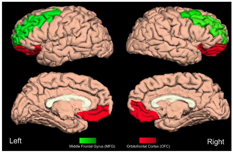
Subjects included 157 patients with neurodegenerative disease. A semiautomated parcellation program (Freesurfer) was used to generate regional cortical volumes from structural MRI scans. Regions of interest (ROIs) included anterior cingulate cortex (ACC), orbitofrontal cortex (OFC), middle frontal gyrus (MFG), and inferior frontal gyrus (IFG). Socioemotional disinhibition was measured using the Neuropsychiatric Inventory. Principal component analysis including 3 tasks of executive function (EF; verbal fluency, Stroop Interference, modified Trails) was used to generate a single-factor score to represent EF.
Partial correlations between ROIs, disinhibition, and EF were computed after controlling for total intracranial volume, Mini-Mental State Examination, diagnosis, age, and education. Brain regions significantly correlated with disinhibition (ACC, OFC, IFG, and temporal lobes) and EF (MFG) were entered into separate hierarchical regressions to determine which brain regions predicted disinhibition and EF. OFC was the only brain region to significantly predict disinhibition, and MFG significantly predicted EF performance. A multivariate general linear model demonstrated a significant interaction between ROIs and cognitive-behavioral functions.
These results support a specific association between orbitofrontal areas and behavioral management as compared with dorsolateral areas and EF.
确定社会情感抑制和执行功能障碍是否与神经退行性疾病的可分离性脑萎缩模式相关。先前的研究表明,神经退行性疾病中的行为和认知功能障碍与额叶不同部位的萎缩有关,但这些先前的研究并未确定这些关系是特定的,这最好通过双重分离来证明。
研究对象包括 157 名神经退行性疾病患者。使用半自动分割程序(Freesurfer)从结构 MRI 扫描生成皮质区域体积。感兴趣区域(ROI)包括前扣带皮层(ACC)、眶额皮层(OFC)、额中回(MFG)和额下回(IFG)。使用神经精神病学问卷测量社会情感抑制。包括 3 项执行功能任务(EF;言语流畅性、Stroop 干扰、改良 Trails)的主成分分析用于生成代表 EF 的单因素得分。
在控制总颅内体积、简易精神状态检查、诊断、年龄和教育后,计算了 ROI、抑制和 EF 之间的部分相关。与抑制(ACC、OFC、IFG 和颞叶)和 EF(MFG)显著相关的脑区被纳入单独的分层回归,以确定哪些脑区预测抑制和 EF。OFC 是唯一显著预测抑制的脑区,MFG 显著预测 EF 表现。多元线性模型显示 ROI 和认知-行为功能之间存在显著的交互作用。
这些结果支持眶额区域与行为管理之间的特定关联,而与背外侧区域和 EF 相关。