Wang Qing, Zhao Guiqiu, Xing Shichao, Zhang Lina, Yang Xian
Department of Ophthalmology, the Affiliated Hospital of Medical College, Qingdao University, Qingdao, China.
Mol Vis. 2011 Mar 8;17:647-57.
To clarify the role of bone morphogenetic proteins (BMP-2,-4,-5) in sclera remodeling during myopia induction and their effect on sclera fibroblasts in cell culture.
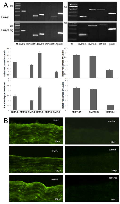
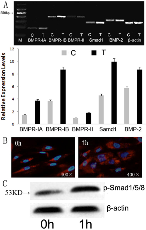
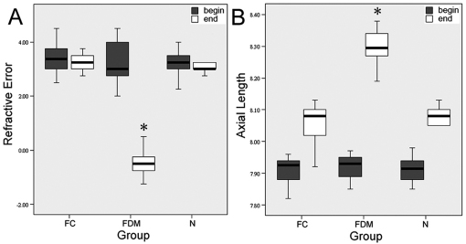
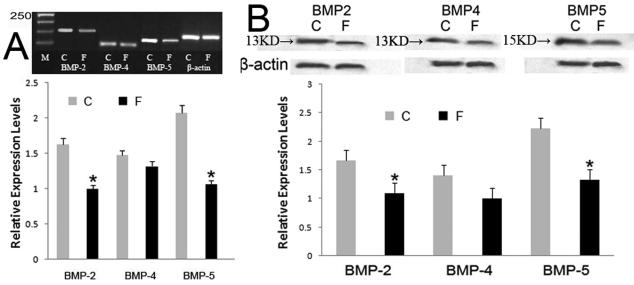
Reverse transcription and polymerase chain reaction (RT-PCR) as well as immunofluorescence were used to detect the expression of the BMPs in human and guinea pig posterior sclera. In guinea pig form-deprivation myopia (FDM) model, RT-PCR and western blotting were used to investigate changes of BMP expression in the posterior sclera. Human sclera fibroblast (HSF) was primarlly cultured and treated with various doses of BMP-2. Cell proliferation was evaluated by the MTT assay. RT-PCR and western-blot were used to determine the changes of collagen I, aggrecan, and possible activated signal pathway. Cell phenotype and activated signal pathway, especially for α-smooth muscle actin (α-SMA) and phospho-smad1/5/8 were then further investigated by cytoimmunofluorescence staining.
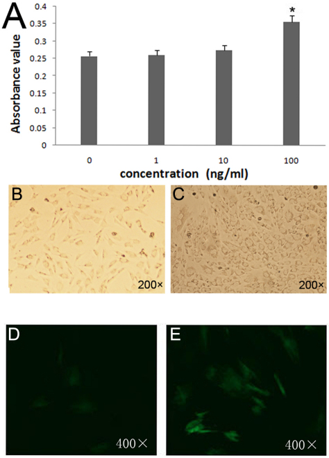
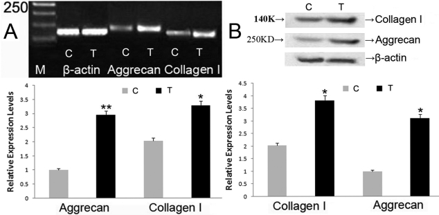
Both human and guinea pig sclera express BMP-2, -4, and -5. In FDM eyes, BMP-2 and BMP-5 expression were reduced in the posterior sclera. Cell proliferation increased significantly (p<0.05) and more cells differentiated into myofibroblast when incubated with 100 ng/ml BMP-2 . The expressions of collangen I, aggrecan, and phospho-smad1/5/8 significantly increased (p<0.05 respectively) as well.
Various BMPs were expressed in human and guinea pig sclera. In the posterior sclera, the expressions of BMP-2 and BMP-5 decreased in FDM eyes. BMP-2 might be able to promote HSF proliferation and differentiation, as well as to help extracellular matrix synthesis potentially through classical Smad pathway.
阐明骨形态发生蛋白(BMP-2、-4、-5)在近视诱导过程中巩膜重塑中的作用及其对细胞培养中巩膜成纤维细胞的影响。
采用逆转录聚合酶链反应(RT-PCR)和免疫荧光法检测人及豚鼠后巩膜中骨形态发生蛋白的表达。在豚鼠形觉剥夺性近视(FDM)模型中,采用RT-PCR和蛋白质印迹法研究后巩膜中骨形态发生蛋白表达的变化。原代培养人巩膜成纤维细胞(HSF),并用不同剂量的BMP-2处理。采用MTT法评估细胞增殖。用RT-PCR和蛋白质印迹法检测Ⅰ型胶原、聚集蛋白聚糖的变化及可能激活的信号通路。然后通过细胞免疫荧光染色进一步研究细胞表型和激活的信号通路,特别是α-平滑肌肌动蛋白(α-SMA)和磷酸化Smad1/5/8。
人及豚鼠巩膜均表达BMP-2、-4和-5。在FDM眼中,后巩膜中BMP-2和BMP-5的表达降低。当用100 ng/ml BMP-2孵育时,细胞增殖显著增加(p<0.05),更多细胞分化为肌成纤维细胞。Ⅰ型胶原、聚集蛋白聚糖和磷酸化Smad1/5/8的表达也显著增加(分别为p<0.05)。
人及豚鼠巩膜中均表达多种骨形态发生蛋白。在FDM眼中,后巩膜中BMP-2和BMP-5的表达降低。BMP-2可能能够促进HSF增殖和分化,并可能通过经典的Smad途径帮助细胞外基质合成。