Biological Physics Group, School of Physics & Astronomy, University of Manchester, Manchester M139PL, UK.
Prog Biophys Mol Biol. 2011 Oct;107(1):156-68. doi: 10.1016/j.pbiomolbio.2011.06.011. Epub 2011 Jul 7.
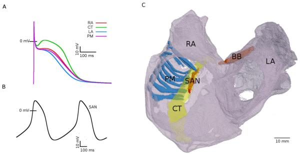
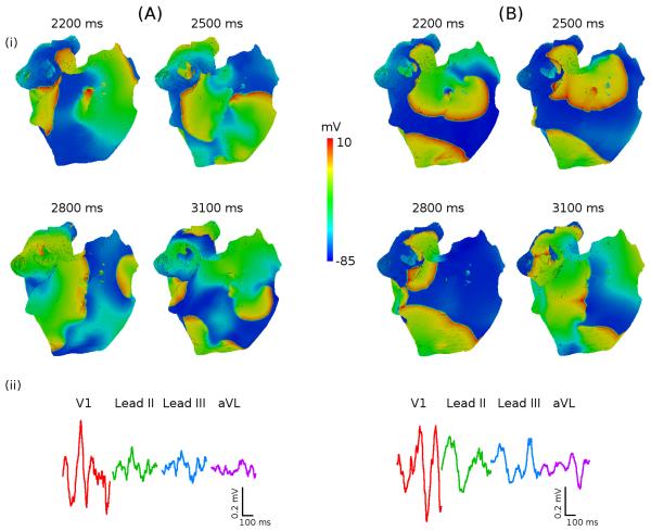
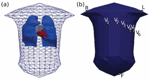
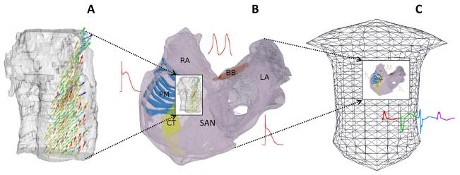
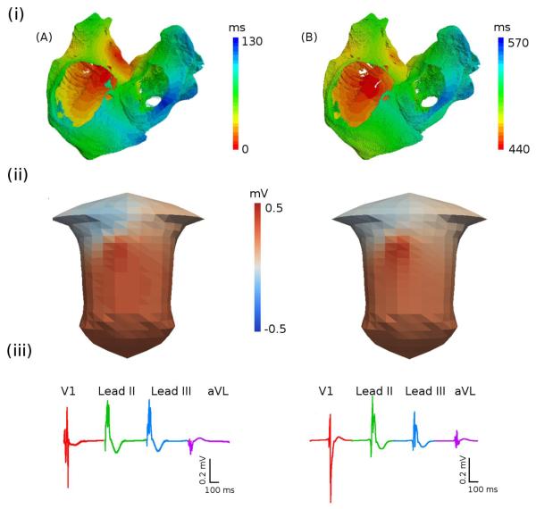
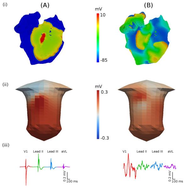
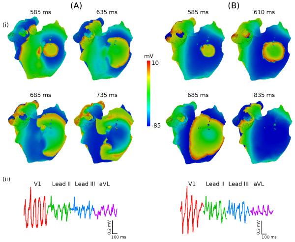
Despite a vast amount of experimental and clinical data on the underlying ionic, cellular and tissue substrates, the mechanisms of common atrial arrhythmias (such as atrial fibrillation, AF) arising from the functional interactions at the whole atria level remain unclear. Computational modelling provides a quantitative framework for integrating such multi-scale data and understanding the arrhythmogenic behaviour that emerges from the collective spatio-temporal dynamics in all parts of the heart. In this study, we have developed a multi-scale hierarchy of biophysically detailed computational models for the human atria--the 3D virtual human atria. Primarily, diffusion tensor MRI reconstruction of the tissue geometry and fibre orientation in the human sinoatrial node (SAN) and surrounding atrial muscle was integrated into the 3D model of the whole atria dissected from the Visible Human dataset. The anatomical models were combined with the heterogeneous atrial action potential (AP) models, and used to simulate the AP conduction in the human atria under various conditions: SAN pacemaking and atrial activation in the normal rhythm, break-down of regular AP wave-fronts during rapid atrial pacing, and the genesis of multiple re-entrant wavelets characteristic of AF. Contributions of different properties of the tissue to mechanisms of the normal rhythm and arrhythmogenesis were investigated. Primarily, the simulations showed that tissue heterogeneity caused the break-down of the normal AP wave-fronts at rapid pacing rates, which initiated a pair of re-entrant spiral waves; and tissue anisotropy resulted in a further break-down of the spiral waves into multiple meandering wavelets characteristic of AF. The 3D virtual atria model itself was incorporated into the torso model to simulate the body surface ECG patterns in the normal and arrhythmic conditions. Therefore, a state-of-the-art computational platform has been developed, which can be used for studying multi-scale electrical phenomena during atrial conduction and AF arrhythmogenesis. Results of such simulations can be directly compared with electrophysiological and endocardial mapping data, as well as clinical ECG recordings. The virtual human atria can provide in-depth insights into 3D excitation propagation processes within atrial walls of a whole heart in vivo, which is beyond the current technical capabilities of experimental or clinical set-ups.
尽管有大量关于离子、细胞和组织基础的实验和临床数据,但源自整个心房水平功能相互作用的常见房性心律失常(如心房颤动,AF)的机制仍不清楚。计算建模为整合此类多尺度数据提供了一个定量框架,并有助于理解从心脏所有部位的集体时空动力学中产生的心律失常行为。在这项研究中,我们为人类心房开发了一个多层次的生物物理详细计算模型——三维虚拟人类心房。首先,对人体窦房结(SAN)和周围心房组织的组织几何形状和纤维方向进行扩散张量 MRI 重建,然后将其整合到从可视人体数据集剖分的整个心房的 3D 模型中。解剖模型与非均匀心房动作电位(AP)模型相结合,用于模拟各种条件下的人类心房中的 AP 传导:SAN 起搏和正常节律下的心房激活、快速心房起搏时规则 AP 波阵面的破裂、以及 AF 特征的多个折返小波的产生。研究了组织不同特性对正常节律和心律失常发生机制的贡献。首先,模拟结果表明,组织异质性导致在快速起搏率下正常 AP 波阵面的破裂,从而引发一对折返螺旋波;组织各向异性导致螺旋波进一步破裂为 AF 特征的多个蜿蜒小波。3D 虚拟心房模型本身被纳入体模,以模拟正常和心律失常条件下的体表心电图模式。因此,开发了一个最先进的计算平台,可用于研究心房传导和 AF 心律失常发生期间的多尺度电现象。此类模拟的结果可以直接与电生理和心内膜标测数据以及临床 ECG 记录进行比较。虚拟人类心房可以深入了解体内整个心脏心房壁内的 3D 兴奋传播过程,这超出了实验或临床设置的当前技术能力。