Department of Biomedical Engineering, University of Iowa, IA 52242, United States.
J Biomech. 2011 Sep 2;44(13):2489-95. doi: 10.1016/j.jbiomech.2011.06.009. Epub 2011 Jul 28.
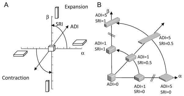
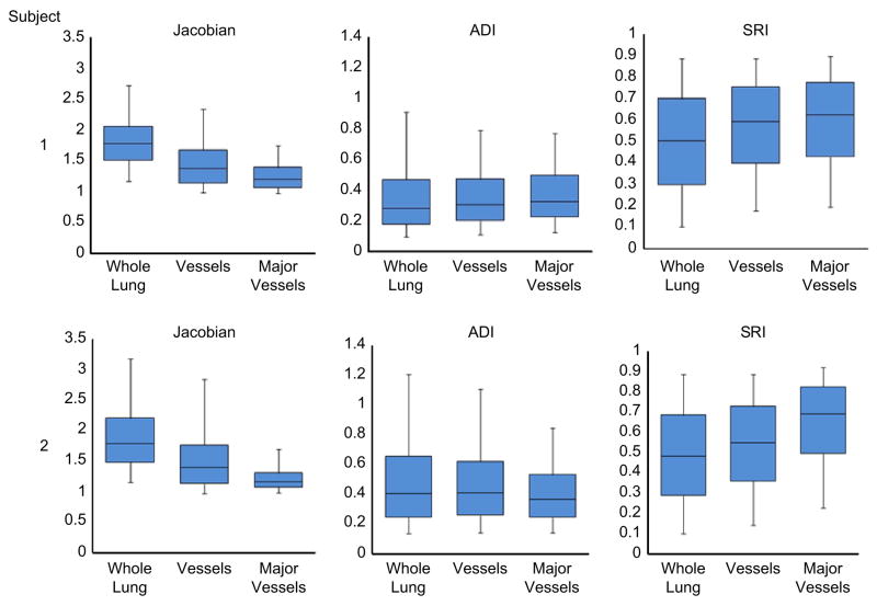
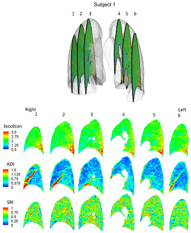
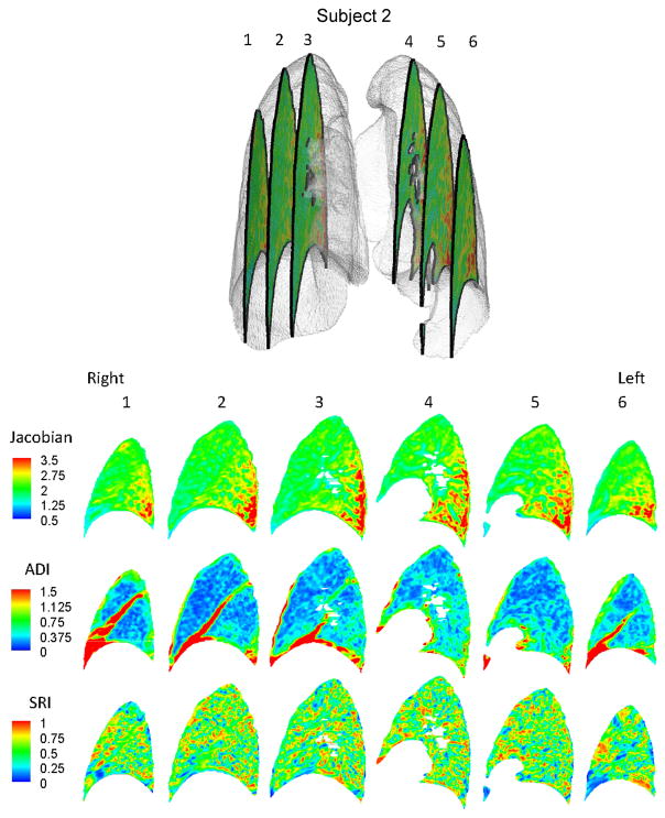
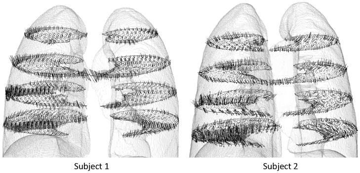
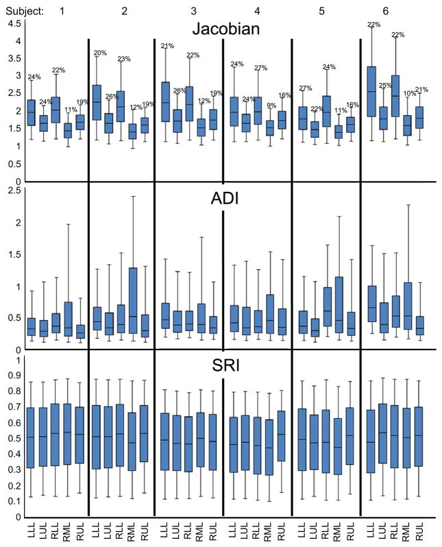
The deformation of the lung during inspiration and expiration involves regional variations in volume change and orientational preferences. Studies have reported techniques for measuring the displacement field in the lung based on imaging or image registration. However, means of interpreting all the information in the displacement field in a physiologically relevant manner is lacking. We propose three indices of lung deformation that are determinable from the displacement field: the Jacobian--a measure of volume change, the anisotropic deformation index--a measure of the magnitude of directional preference in volume change and a slab-rod index--a measure of the nature of directional preference in volume change. To demonstrate the utility of these indices, they were determined for six human subjects using deformable image registration on static CT images, registered from FRC to TLC. Volume change was elevated in the inferior-dorsal region as should be expected for breathing in the supine position. The anisotropic deformation index was elevated in the inferior region owing to proximity to the diaphragm and in the lobar fissures owing to sliding. Vessel regions in the lung had a significantly rod-like deformation compared to the whole lung. Compared to upper lobes, lower lobes exhibited significantly greater volume change (19.4% and 21.3% greater in the right and left lungs on average; p<0.005) and anisotropy in deformation (26.3% and 21.8% greater in the right and left lungs on average; p<0.05) with remarkable consistency across subjects. The developed deformation indices lend themselves to exhaustive and physiologically intuitive interpretations of the displacement fields in the lung determined through image-registration techniques or finite element simulations.
在吸气和呼气过程中,肺部的变形涉及到体积变化的区域变化和定向偏好。研究已经报道了基于成像或图像配准来测量肺部位移场的技术。然而,缺乏以生理相关的方式解释位移场中所有信息的方法。我们提出了三种可从位移场中确定的肺变形指标:雅可比(Jacobian)——衡量体积变化的指标;各向异性变形指数——衡量体积变化方向偏好程度的指标;板杆指数——衡量体积变化方向偏好性质的指标。为了证明这些指标的实用性,我们使用基于静态 CT 图像的可变形图像配准技术,从功能残气位(FRC)到潮气末位(TLC),对六名人类受试者的这些指标进行了确定。正如仰卧位呼吸时所预期的那样,下背部区域的体积变化升高。由于靠近横膈膜和由于滑动,下部区域的各向异性变形指数升高。肺部的血管区域与整个肺部相比具有明显的杆状变形。与上叶相比,下叶的体积变化(右侧和左侧平均增加 19.4%和 21.3%;p<0.005)和变形各向异性(右侧和左侧平均增加 26.3%和 21.8%;p<0.05)显著更大,并且在不同受试者之间具有显著的一致性。所开发的变形指标可以对通过图像配准技术或有限元模拟确定的肺部位移场进行详尽和生理直观的解释。