• 文献检索
  • 文档翻译
  • 深度研究
  • 学术资讯
  • Suppr Zotero 插件Zotero 插件
  • 邀请有礼
  • 套餐&价格
  • 历史记录
应用&插件
Suppr Zotero 插件Zotero 插件浏览器插件Mac 客户端Windows 客户端微信小程序
定价
高级版会员购买积分包购买API积分包
服务
文献检索文档翻译深度研究API 文档MCP 服务
关于我们
关于 Suppr公司介绍联系我们用户协议隐私条款
关注我们

Suppr 超能文献

核心技术专利:CN118964589B侵权必究
粤ICP备2023148730 号-1Suppr @ 2026

文献检索

告别复杂PubMed语法,用中文像聊天一样搜索,搜遍4000万医学文献。AI智能推荐,让科研检索更轻松。

立即免费搜索

文件翻译

保留排版,准确专业,支持PDF/Word/PPT等文件格式,支持 12+语言互译。

免费翻译文档

深度研究

AI帮你快速写综述,25分钟生成高质量综述,智能提取关键信息,辅助科研写作。

立即免费体验

三种谱域光学相干断层扫描系统测量脉络膜厚度的可重复性。

Reproducibility of choroidal thickness measurements across three spectral domain optical coherence tomography systems.

机构信息

New England Eye Center, Tufts Medical Center, Boston, Massachusetts 02111, USA.

出版信息

Ophthalmology. 2012 Jan;119(1):119-23. doi: 10.1016/j.ophtha.2011.07.002. Epub 2011 Sep 23.

DOI:10.1016/j.ophtha.2011.07.002
PMID:21943786
原文链接:https://pmc.ncbi.nlm.nih.gov/articles/PMC3251715/
Abstract

PURPOSE

To investigate the reproducibility of choroidal thickness measurements in normal subjects on 3 spectral domain optical coherence tomography (SD-OCT) instruments: Zeiss Cirrus HD-OCT (Carl Zeiss Meditec Inc., Dublin, CA), Heidelberg Spectralis (Heidelberg Engineering, Heidelberg, Germany), and Optovue RTVue (Optovue Inc., Fremont, CA).

DESIGN

Cross-sectional non-interventional study.

PARTICIPANTS

Images were obtained in 28 eyes of 28 healthy undilated volunteers without ocular pathology in a clinical setting.

METHODS

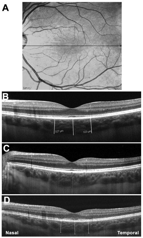
All subjects were imaged on the fovea using Cirrus HD 1-line raster, Spectralis enhanced depth imaging (EDI), and RTVue retina-cross.

MAIN OUTCOME MEASURES

The choroid was measured subfoveally, 750 μm temporal, and 750 μm nasal to the fovea. All measurements were performed by 2 independent observers. Two-way analysis of variance (ANOVA) with Bonferroni's post-test, Pearson correlation, and Bland-Altman analysis were used to compare measurements.

RESULTS

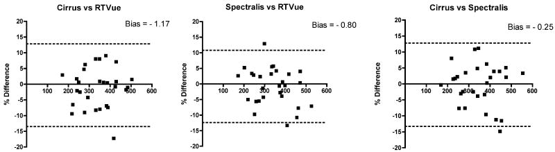
The group of 28 subjects consisted of 7 men and 21 women, with an average age of 35.2 years (range, 23-64 years). A 2-way ANOVA with Bonferroni's post-test revealed no significant difference in the average subfoveal choroidal thickness (P > 0.05) among systems for any location: subfoveally, 750 μm temporal, and 750 μm nasal to the fovea. The measurements of choroidal thickness from any pair of 3 instruments (Cirrus vs. Spectralis, Cirrus vs. RTVue, Spectralis vs. RTVue) were also strongly correlated. The Pearson correlation among all 2 system pairs of the 3 systems was greater than 0.9 (P < 0.0001). The 95% limits of agreement among 4 choroidal thickness measurements were +11.21% to -13.57% (bias -1.17) between Cirrus and RTVue, +10.85% to -12.45% (bias -0.80) between Spectralis and RTVue, and +12.81% to -13.33% (bias -0.25) between Cirrus and Spectralis.

CONCLUSIONS

In our population of young healthy adults with normal vision, there was good reproducibility among choroidal thickness measurements of images acquired with Cirrus, Spectralis, and RTVue.

FINANCIAL DISCLOSURE(S): Proprietary or commercial disclosure may be found after the references.

摘要

目的

研究在 3 种谱域光学相干断层扫描(SD-OCT)仪器(蔡司 Cirrus HD-OCT、海德堡 Spectralis 和 Optovue RTVue)上对正常受试者的脉络膜厚度测量的可重复性。

设计

横断面非干预性研究。

参与者

在临床环境中,在 28 名无眼部病变的健康未散瞳志愿者的 28 只眼中获得图像。

方法

使用 Cirrus HD 1 线光栅、Spectralis 增强深度成像(EDI)和 RTVue 视网膜交叉对所有受试者的黄斑进行成像。

主要观察指标

在黄斑下、750μm 颞侧和黄斑鼻侧进行脉络膜测量。所有测量均由 2 位独立观察者进行。采用双因素方差分析(ANOVA)和 Bonferroni 事后检验、Pearson 相关分析和 Bland-Altman 分析比较测量值。

结果

28 名受试者中包括 7 名男性和 21 名女性,平均年龄 35.2 岁(范围,23-64 岁)。双因素方差分析和 Bonferroni 事后检验显示,在任何位置(黄斑下、750μm 颞侧和黄斑鼻侧),系统之间的平均黄斑下脉络膜厚度均无显著差异(P>0.05)。任何 3 种仪器(Cirrus 与 Spectralis、Cirrus 与 RTVue、Spectralis 与 RTVue)的脉络膜厚度测量结果也具有很强的相关性。3 种系统中所有 2 个系统对的 Pearson 相关系数均大于 0.9(P<0.0001)。Cirrus 和 RTVue 之间的 4 次脉络膜厚度测量的 95%一致性界限为+11.21%至-13.57%(偏倚-1.17),Spectralis 和 RTVue 之间为+10.85%至-12.45%(偏倚-0.80),Cirrus 和 Spectralis 之间为+12.81%至-13.33%(偏倚-0.25)。

结论

在我们的视力正常的年轻健康成年人人群中,Cirrus、Spectralis 和 RTVue 采集的图像的脉络膜厚度测量具有良好的可重复性。

利益冲突

参考文献后可能会发现专有或商业披露。

https://cdn.ncbi.nlm.nih.gov/pmc/blobs/ed32/3251715/4cd89ce5fda8/nihms310956f2.jpg
https://cdn.ncbi.nlm.nih.gov/pmc/blobs/ed32/3251715/3b92a3696c4c/nihms310956f1.jpg
https://cdn.ncbi.nlm.nih.gov/pmc/blobs/ed32/3251715/4cd89ce5fda8/nihms310956f2.jpg
https://cdn.ncbi.nlm.nih.gov/pmc/blobs/ed32/3251715/3b92a3696c4c/nihms310956f1.jpg
https://cdn.ncbi.nlm.nih.gov/pmc/blobs/ed32/3251715/4cd89ce5fda8/nihms310956f2.jpg

相似文献

1
Reproducibility of choroidal thickness measurements across three spectral domain optical coherence tomography systems.三种谱域光学相干断层扫描系统测量脉络膜厚度的可重复性。
Ophthalmology. 2012 Jan;119(1):119-23. doi: 10.1016/j.ophtha.2011.07.002. Epub 2011 Sep 23.
2
Reproducibility of choroidal thickness measurements in subjects on 3 spectral domain optical coherence tomography machines.在3台光谱域光学相干断层扫描机器上对受试者脉络膜厚度测量的可重复性。
Int Ophthalmol. 2017 Jun;37(3):655-671. doi: 10.1007/s10792-016-0306-4. Epub 2016 Aug 24.
3
Macular thickness measurements in healthy eyes using six different optical coherence tomography instruments.使用六种不同光学相干断层扫描仪器对健康眼睛进行黄斑厚度测量。
Invest Ophthalmol Vis Sci. 2009 Jul;50(7):3432-7. doi: 10.1167/iovs.08-2970. Epub 2009 Feb 21.
4
Image inversion spectral-domain optical coherence tomography optimizes choroidal thickness and detail through improved contrast.反转像谱域光学相干断层扫描通过提高对比度来优化脉络膜厚度和细节。
Invest Ophthalmol Vis Sci. 2012 Apr 6;53(4):1874-82. doi: 10.1167/iovs.11-9290.
5
Comparison of foveal microstructure imaging with different spectral domain optical coherence tomography machines.不同谱域光相干断层扫描仪的黄斑区微观结构成像比较。
Ophthalmology. 2012 Nov;119(11):2319-27. doi: 10.1016/j.ophtha.2012.07.012. Epub 2012 Sep 19.
6
Comparison of the diagnostic accuracies of the Spectralis, Cirrus, and RTVue optical coherence tomography devices in glaucoma.比较 Spectralis、Cirrus 和 RTVue 光学相干断层扫描仪在青光眼诊断中的准确性。
Ophthalmology. 2011 Jul;118(7):1334-9. doi: 10.1016/j.ophtha.2010.11.029. Epub 2011 Mar 5.
7
Analysis of choroidal morphologic features and vasculature in healthy eyes using spectral-domain optical coherence tomography.利用频域光学相干断层扫描分析健康眼的脉络膜形态特征和血管。
Ophthalmology. 2013 Sep;120(9):1901-8. doi: 10.1016/j.ophtha.2013.01.066. Epub 2013 May 9.
8
Reproducibility of spectral-domain optical coherence tomography retinal thickness measurements and conversion to equivalent time-domain metrics in diabetic macular edema.糖尿病性黄斑水肿中光谱域光学相干断层扫描视网膜厚度测量的可重复性及向等效时域指标的转换
JAMA Ophthalmol. 2014 Sep;132(9):1113-22. doi: 10.1001/jamaophthalmol.2014.1698.
9
Repeatability and reproducibility of subfoveal choroidal thickness in normal eyes of Japanese using different SD-OCT devices.不同 SD-OCT 设备测量日本人正常眼黄斑中心凹下脉络膜厚度的重复性和可再现性。
Invest Ophthalmol Vis Sci. 2012 Mar 1;53(3):1102-7. doi: 10.1167/iovs.11-8836.
10
Assessment of artifacts and reproducibility across spectral- and time-domain optical coherence tomography devices.跨光谱域和时域光学相干断层扫描设备的伪影评估及可重复性评估
Ophthalmology. 2009 Oct;116(10):1960-70. doi: 10.1016/j.ophtha.2009.03.034. Epub 2009 Jul 9.

引用本文的文献

1
Repeatability, reproducibility, and agreement for three optical coherence tomography instruments.三种光学相干断层扫描仪器的可重复性、再现性及一致性
BMC Ophthalmol. 2025 Jul 28;25(1):427. doi: 10.1186/s12886-025-04257-8.
2
Subfoveal Choroidal Thickness After Successful Retinal Detachment Repair with Persistent Subretinal Fluid at the Macula.黄斑区存在持续性视网膜下液的视网膜脱离修复成功后黄斑中心凹下脉络膜厚度
Clin Ophthalmol. 2025 Jun 11;19:1847-1854. doi: 10.2147/OPTH.S520076. eCollection 2025.
3
Retinal Imaging as a Window into Cardiovascular Health: Towards Harnessing Retinal Analytics for Precision Cardiovascular Medicine.

本文引用的文献

1
Optical coherence tomography enhanced depth imaging of choroidal tumors.光学相干断层扫描增强深度成像在脉络膜肿瘤中的应用。
Am J Ophthalmol. 2011 Apr;151(4):586-593.e2. doi: 10.1016/j.ajo.2010.09.028. Epub 2011 Jan 22.
2
Subfoveal choroidal thickness after treatment of Vogt-Koyanagi-Harada disease.Vogt-Koyanagi-Harada 病治疗后的黄斑中心凹下脉络膜厚度。
Retina. 2011 Mar;31(3):510-7. doi: 10.1097/IAE.0b013e3181eef053.
3
Central serous chorioretinopathy: an update on pathogenesis and treatment.中心性浆液性脉络膜视网膜病变:发病机制和治疗的最新进展。
视网膜成像作为洞察心血管健康的窗口:迈向利用视网膜分析实现精准心血管医学
J Cardiovasc Dev Dis. 2025 Jun 17;12(6):230. doi: 10.3390/jcdd12060230.
4
Characteristics of the retinal and choroidal thicknesses in myopic young adult males using swept-source optical coherence tomography.使用扫频源光学相干断层扫描技术对近视年轻成年男性视网膜和脉络膜厚度特征的研究
PeerJ. 2025 Mar 5;13:e19030. doi: 10.7717/peerj.19030. eCollection 2025.
5
Automatic measurement of choroidal thickness with swept-source optical coherence tomography in chronic Vogt-Koyanagi-Harada disease: 3 years' follow-up.扫频源光学相干断层扫描自动测量慢性葡萄膜大脑炎患者的脉络膜厚度:3年随访
J Ophthalmic Inflamm Infect. 2024 Dec 2;14(1):62. doi: 10.1186/s12348-024-00445-7.
6
Optical Coherence Tomography Study of Choroidal Response to Exercise-Induced Hypertension in Chronic Central Serous Chorioretinopathy.慢性中心性浆液性脉络膜视网膜病变中脉络膜对运动诱发高血压反应的光学相干断层扫描研究
J Clin Med. 2024 Nov 1;13(21):6580. doi: 10.3390/jcm13216580.
7
Comparison of subfoveal choroidal thickness in pre-eclamptic, healthy pregnant, and non-pregnant women.子痫前期孕妇、健康孕妇和非孕妇的黄斑中心凹下脉络膜厚度比较。
Indian J Ophthalmol. 2025 Jan 1;73(Suppl 1):S151-S157. doi: 10.4103/IJO.IJO_767_24. Epub 2024 Oct 25.
8
Automatic fovea detection and choroid segmentation for choroidal thickness assessment in optical coherence tomography.用于光学相干断层扫描中脉络膜厚度评估的自动黄斑中心凹检测和脉络膜分割
Int J Ophthalmol. 2024 Oct 18;17(10):1763-1771. doi: 10.18240/ijo.2024.10.01. eCollection 2024.
9
Pachychoroid pigment epitheliopathy in keratoconic eyes.圆锥角膜眼中的厚脉络膜色素上皮病变
Graefes Arch Clin Exp Ophthalmol. 2025 Jan;263(1):87-95. doi: 10.1007/s00417-024-06612-9. Epub 2024 Aug 30.
10
Comparison of manual and artificial intelligence-automated choroidal thickness segmentation of optical coherence tomography imaging in myopic adults.近视成年人中光学相干断层扫描成像的手动与人工智能自动脉络膜厚度分割比较
Eye Vis (Lond). 2024 Jun 3;11(1):21. doi: 10.1186/s40662-024-00385-2.
Eye (Lond). 2010 Dec;24(12):1743-56. doi: 10.1038/eye.2010.130. Epub 2010 Oct 8.
4
Cirrus HD-OCT high definition imaging is another tool available for visualization of the choroid and provides agreement with the finding that the choroidal thickness is increased in central serous chorioretinopathy in comparison to normal eyes.Cirrus HD-OCT高清成像技术是另一种可用于观察脉络膜的工具,它与中心性浆液性脉络膜视网膜病变患者脉络膜厚度相较于正常眼睛增加这一发现相符。
Retina. 2010 Sep;30(8):1320-1; author reply 1321-2. doi: 10.1097/IAE.0b013e3181e798b1.
5
Choroidal thickness in normal eyes measured using Cirrus HD optical coherence tomography.应用 Cirrus HD 光学相干断层扫描仪测量正常眼的脉络膜厚度。
Am J Ophthalmol. 2010 Sep;150(3):325-329.e1. doi: 10.1016/j.ajo.2010.04.018. Epub 2010 Jun 29.
6
Macular thickness interoperator and intraoperator reproducibility in healthy eyes using 7 optical coherence tomography instruments.使用 7 种光学相干断层扫描仪测量健康眼的黄斑厚度在操作者间和操作者内的可重复性。
Am J Ophthalmol. 2010 Aug;150(2):199-204.e1. doi: 10.1016/j.ajo.2010.03.015. Epub 2010 Jun 8.
7
Subfoveal choroidal thickness after treatment of central serous chorioretinopathy.治疗中心性浆液性脉络膜视网膜病变后脉络膜的中心凹下厚度。
Ophthalmology. 2010 Sep;117(9):1792-9. doi: 10.1016/j.ophtha.2010.01.023. Epub 2010 May 15.
8
Enhanced depth imaging optical coherence tomography of the choroid in central serous chorioretinopathy.中心性浆液性脉络膜视网膜病变脉络膜的增强深度成像光学相干断层扫描
Retina. 2009 Nov-Dec;29(10):1469-73. doi: 10.1097/IAE.0b013e3181be0a83.
9
Enhanced depth imaging optical coherence tomography of the choroid in highly myopic eyes.高度近视眼脉络膜的增强深度成像光学相干断层扫描
Am J Ophthalmol. 2009 Sep;148(3):445-50. doi: 10.1016/j.ajo.2009.04.029. Epub 2009 Jul 9.
10
Macular thickness measurements in healthy eyes using six different optical coherence tomography instruments.使用六种不同光学相干断层扫描仪器对健康眼睛进行黄斑厚度测量。
Invest Ophthalmol Vis Sci. 2009 Jul;50(7):3432-7. doi: 10.1167/iovs.08-2970. Epub 2009 Feb 21.