Department of Pharmacology and Physiology, New Jersey Medical School, University of Medicine and Dentistry, New Jersey, Newark, New Jersey, United States of America.
PLoS One. 2011;6(11):e26976. doi: 10.1371/journal.pone.0026976. Epub 2011 Nov 9.
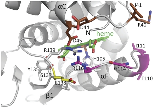
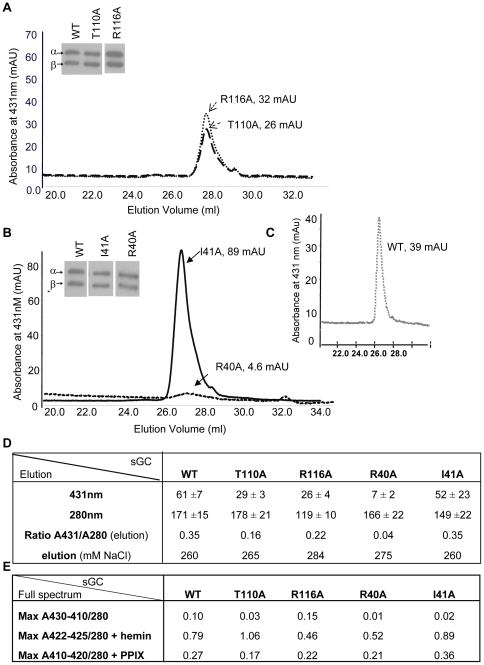
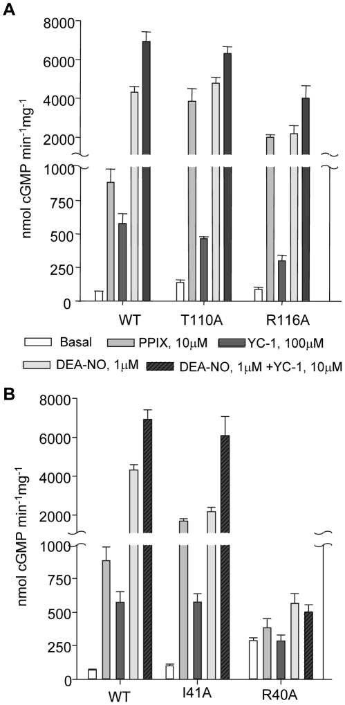
Nitric oxide signals through activation of soluble guanylyl cyclase (sGC), a heme-containing heterodimer. NO binds to the heme domain located in the N-terminal part of the β subunit of sGC resulting in increased production of cGMP in the catalytic domain located at the C-terminal part of sGC. Little is known about the mechanism by which the NO signaling is propagated from the receptor domain (heme domain) to the effector domain (catalytic domain), in particular events subsequent to the breakage of the bond between the heme iron and Histidine 105 (H105) of the β subunit. Our modeling of the heme-binding domain as well as previous homologous heme domain structures in different states point to two regions that could be critical for propagation of the NO activation signal. Structure-based mutational analysis of these regions revealed that residues T110 and R116 in the αF helix-β1 strand, and residues I41 and R40 in the αB-αC loop mediate propagation of activation between the heme domain and the catalytic domain. Biochemical analysis of these heme mutants allows refinement of the map of the residues that are critical for heme stability and propagation of the NO/YC-1 activation signal in sGC.
一氧化氮通过激活可溶性鸟苷酸环化酶(sGC)信号传导,sGC 是一种含有血红素的异二聚体。NO 结合到 sGCβ亚基 N 端部分的血红素结构域,导致位于 sGC C 端部分的催化结构域中 cGMP 的产生增加。关于 NO 信号从受体结构域(血红素结构域)传播到效应结构域(催化结构域)的机制知之甚少,特别是血红素铁与 sGCβ亚基的组氨酸 105(H105)之间的键断裂后的事件。我们对血红素结合结构域的建模以及不同状态下先前同源血红素结构域结构表明,有两个区域可能对 NO 激活信号的传播至关重要。对这些区域进行基于结构的突变分析表明,αF 螺旋-β1 链上的残基 T110 和 R116,以及αB-αC 环上的残基 I41 和 R40 介导血红素结构域和催化结构域之间的激活传播。对这些血红素突变体的生化分析允许细化血红素稳定性和 sGC 中 NO/YC-1 激活信号传播的关键残基图谱。