• 文献检索
  • 文档翻译
  • 深度研究
  • 学术资讯
  • Suppr Zotero 插件Zotero 插件
  • 邀请有礼
  • 套餐&价格
  • 历史记录
应用&插件
Suppr Zotero 插件Zotero 插件浏览器插件Mac 客户端Windows 客户端微信小程序
定价
高级版会员购买积分包购买API积分包
服务
文献检索文档翻译深度研究API 文档MCP 服务
关于我们
关于 Suppr公司介绍联系我们用户协议隐私条款
关注我们

Suppr 超能文献

核心技术专利:CN118964589B侵权必究
粤ICP备2023148730 号-1Suppr @ 2026

文献检索

告别复杂PubMed语法,用中文像聊天一样搜索,搜遍4000万医学文献。AI智能推荐,让科研检索更轻松。

立即免费搜索

文件翻译

保留排版,准确专业,支持PDF/Word/PPT等文件格式,支持 12+语言互译。

免费翻译文档

深度研究

AI帮你快速写综述,25分钟生成高质量综述,智能提取关键信息,辅助科研写作。

立即免费体验

使用光学相干断层扫描和自适应光学对外侧视网膜进行相敏成像。

Phase-sensitive imaging of the outer retina using optical coherence tomography and adaptive optics.

作者信息

Jonnal Ravi S, Kocaoglu Omer P, Wang Qiang, Lee Sangyeol, Miller Donald T

机构信息

Program in Vision Science, Indiana University, 800 East Atwater Avenue, Bloomington, IN 47405, USA.

出版信息

Biomed Opt Express. 2012 Jan 1;3(1):104-24. doi: 10.1364/BOE.3.000104. Epub 2011 Dec 13.

DOI:10.1364/BOE.3.000104
PMID:22254172
原文链接:https://pmc.ncbi.nlm.nih.gov/articles/PMC3255329/
Abstract

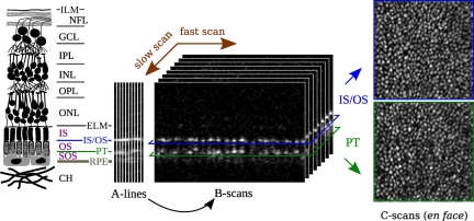
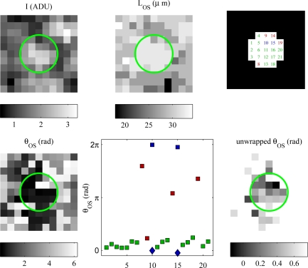
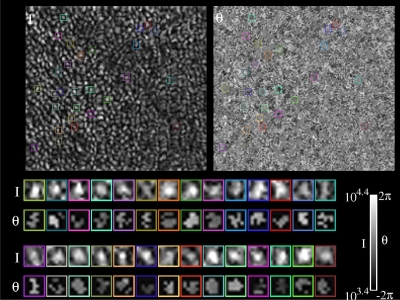
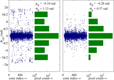
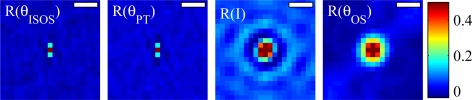
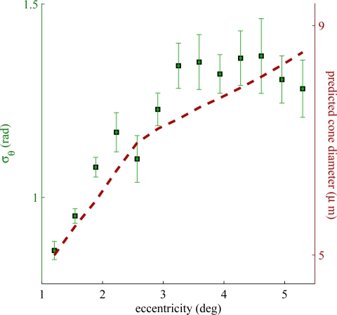
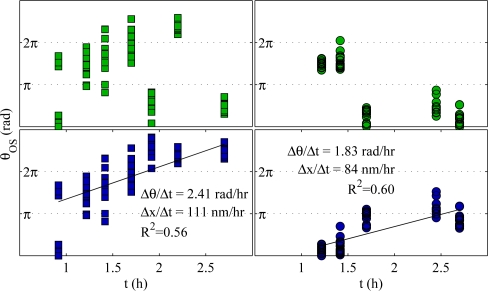
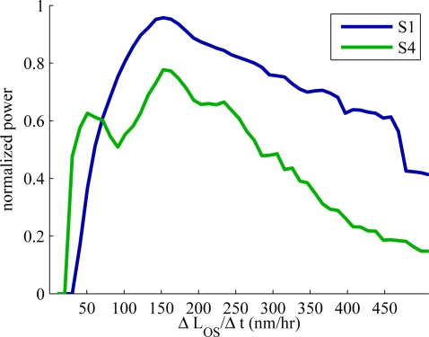
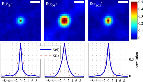
The cone photoreceptor's outer segment (OS) experiences changes in optical path length, both in response to visible stimuli and as a matter of its daily course of renewal and shedding. These changes are of interest, to quantify function in healthy cells and assess dysfunction in diseased ones. While optical coherence tomography (OCT), combined with adaptive optics (AO), has permitted unprecedented three-dimensional resolution in the living retina, it has not generally been able to measure these OS dynamics, whose scale is smaller than OCT's axial resolution of a few microns. A possible solution is to take advantage of the phase information encoded in the OCT signal. Phase-sensitive implementations of spectral-domain optical coherence tomography (SD-OCT) have been demonstrated, capable of resolving sample axial displacements much smaller than the imaging wavelength, but these have been limited to ex vivo samples. In this paper we present a novel technique for retrieving phase information from OCT volumes of the outer retina. The key component of our technique is quantification of phase differences within the retina. We provide a quantitative analysis of such phase information and show that-when combined with appropriate methods for filtering and unwrapping-it can improve the sensitivity to OS length change by more than an order of magnitude, down to 45 nm, slightly thicker than a single OS disc. We further show that phase sensitivity drops off with retinal eccentricity, and that the best location for phase imaging is close to the fovea. We apply the technique to the measurement of sub-resolution changes in the OS over matters of hours. Using custom software for registration and tracking, these microscopic changes are monitored in hundreds of cones over time. In two subjects, the OS was found to have average elongation rates of 150 nm/hr, values which agree with our previous findings.

摘要

视锥光感受器的外段(OS)在响应可见刺激以及日常更新和脱落过程中,其光程长度都会发生变化。这些变化对于量化健康细胞的功能以及评估患病细胞的功能障碍具有重要意义。虽然光学相干断层扫描(OCT)结合自适应光学(AO)技术在活体视网膜中实现了前所未有的三维分辨率,但通常无法测量这些OS动态变化,因为其尺度小于OCT几微米的轴向分辨率。一种可能的解决方案是利用OCT信号中编码的相位信息。光谱域光学相干断层扫描(SD - OCT)的相敏实现方式已得到证实,能够分辨比成像波长小得多的样本轴向位移,但这些仅限于离体样本。在本文中,我们提出了一种从视网膜外层的OCT体积中检索相位信息的新技术。我们技术的关键组成部分是对视网膜内相位差的量化。我们对这种相位信息进行了定量分析,并表明当与适当的滤波和解缠方法相结合时,它可以将对OS长度变化的灵敏度提高一个数量级以上,低至45纳米,略厚于单个OS盘。我们进一步表明,相位灵敏度随视网膜偏心率下降,并且相位成像的最佳位置靠近中央凹。我们将该技术应用于测量数小时内OS的亚分辨率变化。使用定制的配准和跟踪软件,随着时间的推移对数百个视锥细胞中的这些微观变化进行监测。在两名受试者中,发现OS的平均伸长率为150纳米/小时,这一数值与我们之前的研究结果一致。

https://cdn.ncbi.nlm.nih.gov/pmc/blobs/2e8e/3255329/2f71ba8a38ab/boe-3-1-104-g010.jpg
https://cdn.ncbi.nlm.nih.gov/pmc/blobs/2e8e/3255329/828067103a76/boe-3-1-104-g001.jpg
https://cdn.ncbi.nlm.nih.gov/pmc/blobs/2e8e/3255329/3fa41ff618b4/boe-3-1-104-g002.jpg
https://cdn.ncbi.nlm.nih.gov/pmc/blobs/2e8e/3255329/101ec63b95fa/boe-3-1-104-g003.jpg
https://cdn.ncbi.nlm.nih.gov/pmc/blobs/2e8e/3255329/3be72f1d0915/boe-3-1-104-g004.jpg
https://cdn.ncbi.nlm.nih.gov/pmc/blobs/2e8e/3255329/e5c9de6c4ae2/boe-3-1-104-g005.jpg
https://cdn.ncbi.nlm.nih.gov/pmc/blobs/2e8e/3255329/9cfc698f45e9/boe-3-1-104-g006.jpg
https://cdn.ncbi.nlm.nih.gov/pmc/blobs/2e8e/3255329/42d2ecc30c4a/boe-3-1-104-g007.jpg
https://cdn.ncbi.nlm.nih.gov/pmc/blobs/2e8e/3255329/6c96d565205f/boe-3-1-104-g008.jpg
https://cdn.ncbi.nlm.nih.gov/pmc/blobs/2e8e/3255329/87fc646ebc98/boe-3-1-104-g009.jpg
https://cdn.ncbi.nlm.nih.gov/pmc/blobs/2e8e/3255329/2f71ba8a38ab/boe-3-1-104-g010.jpg

相似文献

1
Phase-sensitive imaging of the outer retina using optical coherence tomography and adaptive optics.使用光学相干断层扫描和自适应光学对外侧视网膜进行相敏成像。
Biomed Opt Express. 2012 Jan 1;3(1):104-24. doi: 10.1364/BOE.3.000104. Epub 2011 Dec 13.
2
Imaging outer segment renewal in living human cone photoreceptors.活体人类视锥光感受器中外段更新的成像
Opt Express. 2010 Mar 1;18(5):5257-70. doi: 10.1364/OE.18.005257.
3
Morphologic and Functional Assessment of Photoreceptors After Macula-Off Retinal Detachment With Adaptive-Optics OCT and Microperimetry.黄斑裂孔性视网膜脱离后应用自适应光学 OCT 和微视野计对光感受器的形态和功能评估。
Am J Ophthalmol. 2020 Jun;214:72-85. doi: 10.1016/j.ajo.2019.12.015. Epub 2019 Dec 25.
4
Three-dimensional composition of the photoreceptor cone layers in healthy eyes using adaptive-optics optical coherence tomography (AO-OCT).利用自适应光学光相干断层扫描(AO-OCT)技术对健康眼的光感受器锥体细胞层进行三维成像。
PLoS One. 2021 Jan 7;16(1):e0245293. doi: 10.1371/journal.pone.0245293. eCollection 2021.
5
Imaging cone photoreceptors in three dimensions and in time using ultrahigh resolution optical coherence tomography with adaptive optics.使用具有自适应光学的超高分辨率光学相干断层扫描在三维空间和时间上对视锥光感受器进行成像。
Biomed Opt Express. 2011 Mar 1;2(4):748-63. doi: 10.1364/BOE.2.000748.
6
The fundus photo has met its match: optical coherence tomography and adaptive optics ophthalmoscopy are here to stay.眼底照片遇到了对手:光学相干断层扫描和自适应光学检眼镜将继续存在。
Ophthalmic Physiol Opt. 2016 May;36(3):218-39. doi: 10.1111/opo.12289.
7
Integrity of the cone photoreceptor mosaic in oligocone trichromacy.寡色视锥症中视锥细胞马赛克的完整性。
Invest Ophthalmol Vis Sci. 2011 Jul 1;52(7):4757-64. doi: 10.1167/iovs.10-6659.
8
Photoreceptor disc shedding in the living human eye.活体人眼中光感受器盘膜的脱落。
Biomed Opt Express. 2016 Oct 13;7(11):4554-4568. doi: 10.1364/BOE.7.004554. eCollection 2016 Nov 1.
9
Toward a clinical optoretinogram: a review of noninvasive, optical tests of retinal neural function.迈向临床视网膜电图:视网膜神经功能的非侵入性光学检测综述
Ann Transl Med. 2021 Aug;9(15):1270. doi: 10.21037/atm-20-6440.
10
Volumetric imaging of rod and cone photoreceptor structure with a combined adaptive optics-optical coherence tomography-scanning laser ophthalmoscope.利用自适应光学-光学相干断层扫描-扫描激光检眼镜对杆状和锥状光感受器结构进行容积成像。
J Biomed Opt. 2018 Mar;23(3):1-15. doi: 10.1117/1.JBO.23.3.036003.

引用本文的文献

1
Optoretinography with actively stabilized adaptive optics optical coherence tomography.采用主动稳定自适应光学光学相干断层扫描技术的视网膜电图检查。
Biomed Opt Express. 2025 Jul 17;16(8):3222-3236. doi: 10.1364/BOE.566376. eCollection 2025 Aug 1.
2
Insight into human photoreceptor function: Modeling optoretinographic responses to diverse stimuli.深入了解人类光感受器功能:模拟视网膜电图对各种刺激的反应。
Sci Adv. 2025 Jun 13;11(24):eadq7332. doi: 10.1126/sciadv.adq7332. Epub 2025 Jun 11.
3
Velocity-based optoretinography for clinical applications.

本文引用的文献

1
Imaging single cells in the living retina.对活体视网膜中的单细胞进行成像。
Vision Res. 2011 Jul 1;51(13):1379-96. doi: 10.1016/j.visres.2011.05.002. Epub 2011 May 10.
2
Imaging cone photoreceptors in three dimensions and in time using ultrahigh resolution optical coherence tomography with adaptive optics.使用具有自适应光学的超高分辨率光学相干断层扫描在三维空间和时间上对视锥光感受器进行成像。
Biomed Opt Express. 2011 Mar 1;2(4):748-63. doi: 10.1364/BOE.2.000748.
3
Temporal changes of human cone photoreceptors observed in vivo with SLO/OCT.
用于临床应用的基于速度的视网膜电图
Optica. 2022 Oct 20;9(10):1100-1108. doi: 10.1364/optica.460835. Epub 2022 Sep 22.
4
Insight into human photoreceptor function: modeling optoretinographic responses to diverse stimuli.深入了解人类光感受器功能:模拟视网膜电图对不同刺激的反应。
bioRxiv. 2025 Feb 28:2025.02.28.639986. doi: 10.1101/2025.02.28.639986.
5
Phase-restoring subpixel image registration: enhancing motion detection performance in Fourier-domain optical coherence tomography.相位恢复亚像素图像配准:提高傅里叶域光学相干断层扫描中的运动检测性能
J Phys D Appl Phys. 2025 Apr 7;58(14):145102. doi: 10.1088/1361-6463/adb3b4. Epub 2025 Feb 21.
6
From Cellular to Metabolic: Advances in Imaging of Inherited Retinal Diseases.从细胞层面到代谢层面:遗传性视网膜疾病成像技术的进展
Diagnostics (Basel). 2024 Dec 26;15(1):28. doi: 10.3390/diagnostics15010028.
7
Intensity-based optoretinography reveals sub-clinical deficits in cone function in retinitis pigmentosa.基于强度的视网膜电图揭示了色素性视网膜炎患者视锥细胞功能的亚临床缺陷。
Front Ophthalmol (Lausanne). 2024 Jun 4;4:1373549. doi: 10.3389/fopht.2024.1373549. eCollection 2024.
8
Evolution of adaptive optics retinal imaging [Invited].自适应光学视网膜成像的发展[特邀报告]
Biomed Opt Express. 2023 Feb 28;14(3):1307-1338. doi: 10.1364/BOE.485371. eCollection 2023 Mar 1.
9
Coarse-scale optoretinography (CoORG) with extended field-of-view for normative characterization.用于规范表征的具有扩展视野的粗尺度视网膜电图(CoORG)。
Biomed Opt Express. 2022 Oct 24;13(11):5989-6002. doi: 10.1364/BOE.473475. eCollection 2022 Nov 1.
10
Retinal imaging using adaptive optics optical coherence tomography with fast and accurate real-time tracking.使用自适应光学光学相干断层扫描并具备快速准确实时跟踪功能的视网膜成像。
Biomed Opt Express. 2022 Oct 18;13(11):5909-5925. doi: 10.1364/BOE.467634. eCollection 2022 Nov 1.
利用扫描激光检眼镜/光学相干断层扫描技术在体内观察到的人类视锥光感受器的时间变化。
Biomed Opt Express. 2010 Dec 15;2(1):100-12. doi: 10.1364/BOE.2.000100.
4
Imaging outer segment renewal in living human cone photoreceptors.活体人类视锥光感受器中外段更新的成像
Opt Express. 2010 Mar 1;18(5):5257-70. doi: 10.1364/OE.18.005257.
5
Retinal imaging with polarization-sensitive optical coherence tomography and adaptive optics.使用偏振敏感光学相干断层扫描和自适应光学技术的视网膜成像。
Opt Express. 2009 Nov 23;17(24):21634-51. doi: 10.1364/OE.17.021634.
6
In vivo functional imaging of human cone photoreceptors.人类视锥光感受器的体内功能成像。
Opt Express. 2007;15(4):16141-16160. doi: 10.1364/OE.15.016141.
7
Optical coherence angiography.光学相干血管造影术。
Opt Express. 2006 Aug 21;14(17):7821-40. doi: 10.1364/oe.14.007821.
8
Adaptive optics parallel spectral domain optical coherence tomography for imaging the living retina.用于活体视网膜成像的自适应光学并行光谱域光学相干断层扫描技术
Opt Express. 2005 Jun 13;13(12):4792-811. doi: 10.1364/opex.13.004792.
9
Detection of neural activity using phase-sensitive optical low-coherence reflectometry.使用相敏光学低相干反射测量法检测神经活动。
Opt Express. 2004 May 31;12(11):2377-86. doi: 10.1364/opex.12.002377.
10
Real-time assessment of retinal blood flow with ultrafast acquisition by color Doppler Fourier domain optical coherence tomography.通过彩色多普勒傅里叶域光学相干断层扫描进行超快速采集对视网膜血流进行实时评估。
Opt Express. 2003 Nov 17;11(23):3116-21. doi: 10.1364/oe.11.003116.