Geisbert T W, Jahrling P B
Disease Assessment Division, United States Army Medical Research Institute of Infectious Diseases, Fort Detrick, Frederick, Maryland 21701-5011.
J Clin Pathol. 1990 Oct;43(10):813-6. doi: 10.1136/jcp.43.10.813.
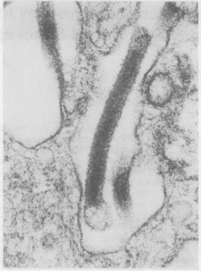
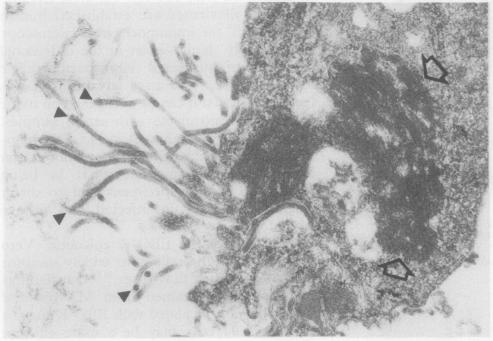
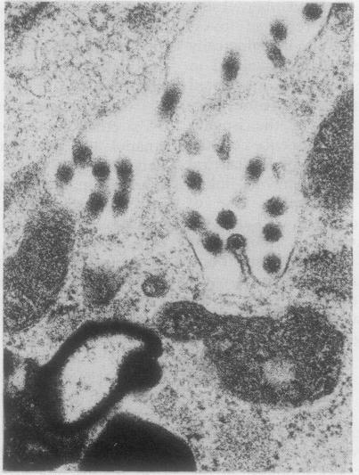
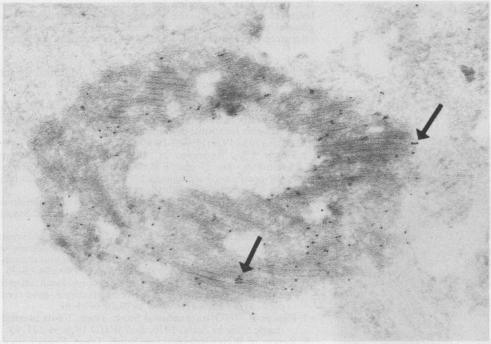
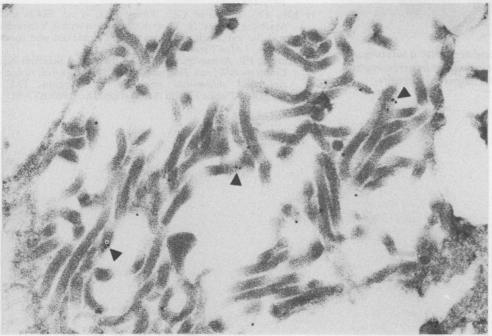
A filovirus, serologically related to Ebola virus, was detected by "post-embedment" immunoelectron microscopical examination of MA-104 cells. These had been infected by inoculation with serum samples obtained during the 1989 epizootic in cynomolgus monkeys (Macaca fascicularis), imported from the Philippines and maintained at Reston, Virginia, USA, a primate holding facility. The immunoelectron microscopy method, when used in conjunction with standard transmission electron microscopy (TEM) of infected cells, provided consistent results and was simple to perform in this epizootic. It is concluded that immunoelectron microscopy is potentially useful in the direct immunological diagnosis of Ebola and related filoviral infections (such as Marburg) in clinical samples obtained from those with acute infection.
通过对MA - 104细胞进行“包埋后”免疫电子显微镜检查,检测到一种与埃博拉病毒血清学相关的丝状病毒。这些细胞是用从菲律宾进口并饲养在美国弗吉尼亚州雷斯顿的灵长类动物饲养设施中的食蟹猴(猕猴)在1989年 epizootic期间获得的血清样本接种感染的。免疫电子显微镜方法与感染细胞的标准透射电子显微镜(TEM)结合使用时,能提供一致的结果,并且在这次 epizootic中操作简单。得出的结论是,免疫电子显微镜在从急性感染患者获得的临床样本中对埃博拉及相关丝状病毒感染(如马尔堡)的直接免疫诊断中可能有用。