Lin A, Krockmalnic G, Penman S
Department of Biology, Massachusetts Institute of Technology, Cambridge 02139.
Proc Natl Acad Sci U S A. 1990 Nov;87(21):8565-9. doi: 10.1073/pnas.87.21.8565.
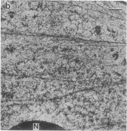
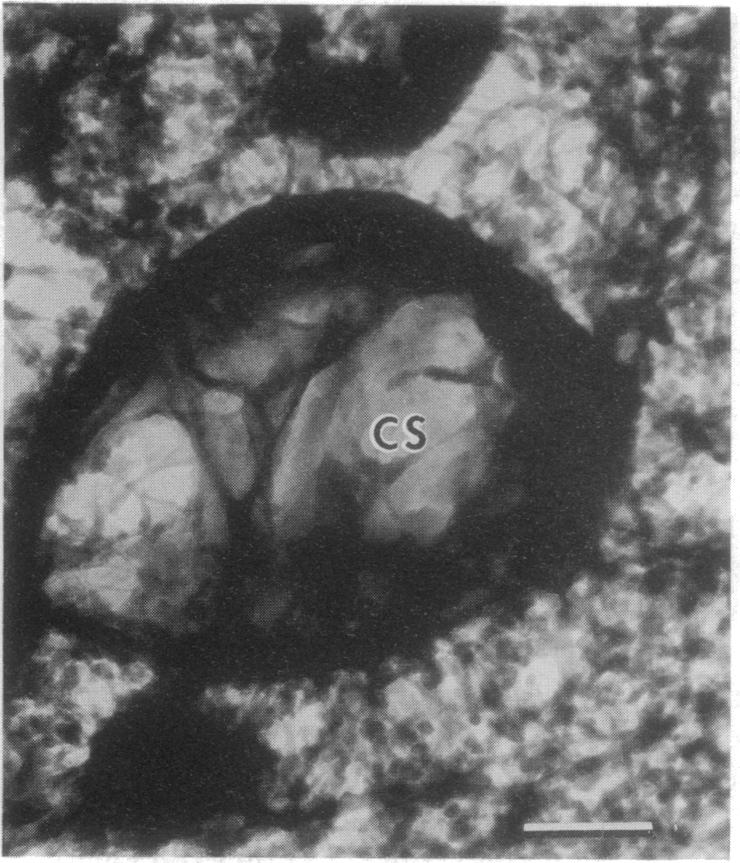
Embedment-free electron microscopy images the cytoskeleton and nuclear matrix, which are very difficult to visualize in conventional electron micrographs. However, to be effective, cell structures must be depleted of soluble proteins, which otherwise shroud cell architecture. Nonionic detergents effect this extraction, releasing soluble proteins but also destroying all membranes. Saponin can permeabilize plasma membranes, releasing soluble proteins while preserving many cytoplasmic membranes. Stereoscopic electron microscopy of resinless sections shows the many connections of the cytoskeleton to mitochondrial membranes.
无包埋电子显微镜可对细胞骨架和核基质进行成像,而在传统电子显微照片中很难看到这些结构。然而,为了使成像有效,必须去除细胞结构中的可溶性蛋白质,否则这些蛋白质会掩盖细胞结构。非离子去污剂可实现这种提取,释放可溶性蛋白质,但也会破坏所有膜结构。皂苷可使质膜通透,释放可溶性蛋白质,同时保留许多细胞质膜。对无树脂切片进行立体电子显微镜观察,可显示细胞骨架与线粒体膜之间的许多连接。