Department of Laboratory Medicine, School of Medicine, The Catholic University of Korea, Seoul, Korea.
Ann Lab Med. 2012 May;32(3):171-6. doi: 10.3343/alm.2012.32.3.171. Epub 2012 Apr 18.
We developed a single-color multitarget flow cytometry (SM-FC) assay, a single-tube assay with graded mean fluorescence intensities (MFIs). We evaluated the repeatability of SM-FC, and its correlation with multicolor flow cytometry (MFC), to assess its application as a routine FC assay.
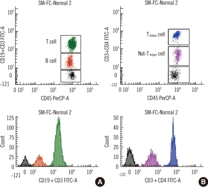
We selected CD19, CD3, CD4, and CD8 as antigen targets to analyze a lymphocyte subset. MFIs were graded by adjusting monoclonal antibody (mAb) volumes to detect several cell populations. Dimly labeled mAb was prepared by decreasing mAb volume and the optimum diluted volume was determined by serial dilution. SM-FC repeatability was analyzed 10 times in 2 normal controls. The correlation between SM-FC and MFC was evaluated in 20 normal and 23 patient samples.
CV values (0.8-5.0% and 1.3-4.1% in samples 1 and 2, respectively) acquired by SM-FC with CD3-fluorescein α-isothyocyanate (FITC)(dim)+CD4-FITC(bright) and with CD19-FITC(dim)+CD3-FITC(bright) showed good repeatability, comparable to that acquired by MFC (1.6-3.7% and 1.0-4.8% in samples 1 and 2, respectively). Excellent correlation was observed between the 2 methods in the 20 normal samples (B cells, T cells, non-T(helper) cells, and T(helper) cells; r(2)=0.87, 0.97, 0.97, and 0.98, respectively; P<0.05). There were also linear relationships between SM-FC with CD19-FITC(dim)+CD3-FITC(bright) and CD8-PE(dim)+CD4-PE(bright), and MFC, in the 23 patient samples (B cells, T cells, T(cytotoxic) cells, and T(helper) cells; r(2)≥0.98, 0.99, 0.99, and 0.99, respectively; P<0.05).
The multicolor, single-tube SM-FC technique is a potential alternative tool for identifying a lymphocyte subset.
我们开发了一种单色彩多靶流式细胞术(SM-FC)检测法,这是一种具有梯度平均荧光强度(MFI)的单管检测法。我们评估了 SM-FC 的重复性及其与多色流式细胞术(MFC)的相关性,以评估其作为常规 FC 检测法的应用。
我们选择 CD19、CD3、CD4 和 CD8 作为抗原靶标来分析淋巴细胞亚群。通过调整单克隆抗体(mAb)体积来检测多个细胞群,对 MFI 进行分级。通过减少 mAb 体积制备暗淡标记的 mAb,并通过连续稀释确定最佳稀释体积。在 2 个正常对照中,10 次分析 SM-FC 重复性。在 20 个正常样本和 23 个患者样本中评估 SM-FC 与 MFC 的相关性。
SM-FC 检测 CD3-荧光素 α-异硫氰酸酯(FITC)(暗淡)+CD4-FITC(明亮)和 CD19-FITC(暗淡)+CD3-FITC(明亮)的 CV 值(样本 1 为 0.8-5.0%,样本 2 为 1.3-4.1%)显示出良好的重复性,与 MFC 获得的重复性相当(样本 1 分别为 1.6-3.7%和 1.0-4.8%,样本 2 分别为 1.6-3.7%和 1.0-4.8%)。在 20 个正常样本中,两种方法之间观察到极好的相关性(B 细胞、T 细胞、非 T(辅助)细胞和 T(辅助)细胞;r(2)分别为 0.87、0.97、0.97 和 0.98;P<0.05)。在 23 个患者样本中,SM-FC 检测 CD19-FITC(暗淡)+CD3-FITC(明亮)与 CD8-PE(暗淡)+CD4-PE(明亮)和 MFC 之间也存在线性关系(B 细胞、T 细胞、T(细胞毒性)细胞和 T(辅助)细胞;r(2)≥0.98、0.99、0.99 和 0.99;P<0.05)。
多色单管 SM-FC 技术是鉴定淋巴细胞亚群的潜在替代工具。