Cadotte David W, Mariampillai Adrian, Cadotte Adam, Lee Kenneth K C, Kiehl Tim-Rasmus, Wilson Brian C, Fehlings Michael G, Yang Victor X D
Biomed Opt Express. 2012 May 1;3(5):911-9. doi: 10.1364/BOE.3.000911. Epub 2012 Apr 10.
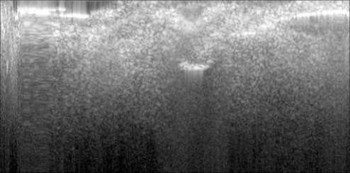
Optical coherence tomography (OCT) has the combined advantage of high temporal (µsec) and spatial (<10µm) resolution. These features make it an attractive tool to study the dynamic relationship between neural activity and the surrounding blood vessels in the spinal cord, a topic that is poorly understood. Here we present work that aims to optimize an in vivo OCT imaging model of the rodent spinal cord. In this study we image the microvascular networks of both rats and mice using speckle variance OCT. This is the first report of depth resolved imaging of the in vivo spinal cord using an entirely endogenous contrast mechanism.
光学相干断层扫描(OCT)具有高时间(微秒)和空间(<10微米)分辨率的综合优势。这些特性使其成为研究脊髓中神经活动与周围血管之间动态关系的一个有吸引力的工具,而这一主题目前了解甚少。在此,我们展示了旨在优化啮齿动物脊髓体内OCT成像模型的工作。在本研究中,我们使用斑点方差OCT对大鼠和小鼠的微血管网络进行成像。这是首次使用完全内源性对比机制对体内脊髓进行深度分辨成像的报告。