Menzies Research Institute Tasmania, 17 Liverpool Street, Hobart, TAS, 7001, Australia.
J Neuroinflammation. 2012 May 29;9:109. doi: 10.1186/1742-2094-9-109.
The primary olfactory pathway is a potential route through which microorganisms from the periphery could potentially access the central nervous system. Our previous studies demonstrated that if the olfactory epithelium was damaged, bacteria administered into the nasal cavity induced nitric oxide production in olfactory ensheathing cells. This study investigates the cytokine profile of olfactory tissues as a consequence of bacterial challenge and establishes whether or not the bacteria are able to reach the olfactory bulb in the central nervous system.
The olfactory epithelium of C57BL/6 mice was damaged by unilateral Triton X-100 nasal washing, and Staphylococcus aureus was administered ipsilaterally 4 days later. Olfactory mucosa and bulb were harvested 6 h, 24 h and 5 days after inoculation and their cytokine profile compared to control tissues. The fate of S. aureus and the response of bulbar microglia were examined using fluorescence microscopy and transmission electron microscopy.
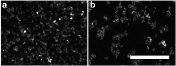
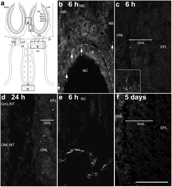
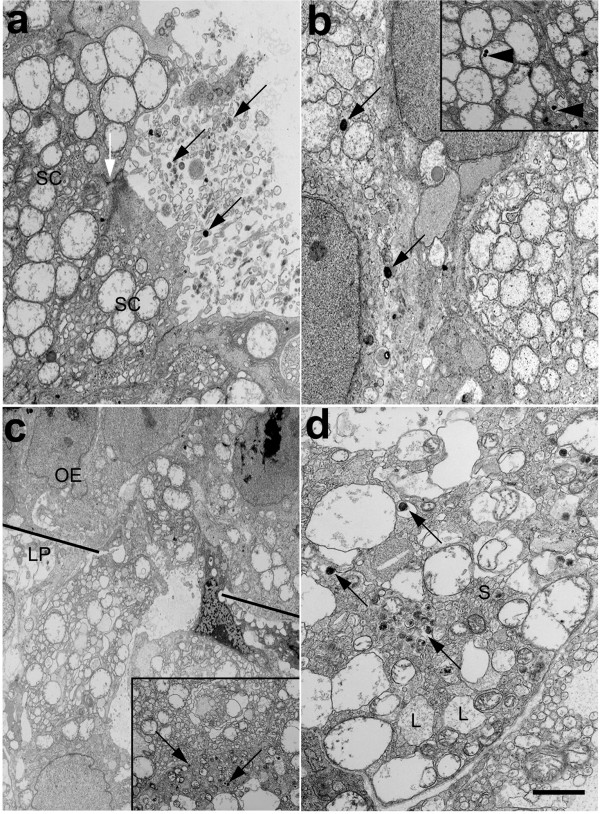
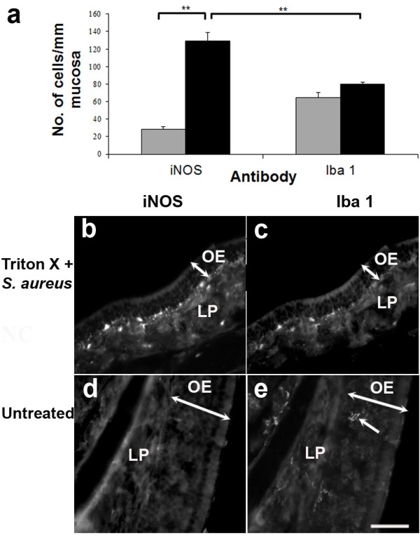
In the olfactory mucosa, administered S. aureus was present in supporting cells of the olfactory epithelium, and macrophages and olfactory nerve bundles in the lamina propria. Fluorescein isothiocyanate-conjugated S. aureus was observed within the olfactory mucosa and bulb 6 h after inoculation, but remained restricted to the peripheral layers up to 5 days later. At the 24-h time point, the level of interleukin-6 (IL-6) and tumour necrosis factor-α in the compromised olfactory tissues challenged with bacteria (12,466 ± 956 pg/ml and 552 ± 193 pg/ml, respectively) was significantly higher than that in compromised olfactory tissues alone (6,092 ± 1,403 pg/ml and 80 ± 2 pg/ml, respectively). Immunohistochemistry confirmed that IL-6 was present in several cell types including olfactory ensheathing cells and mitral cells of the olfactory bulb. Concurrently, there was a 4.4-, 4.5- and 2.8-fold increase in the density of iNOS-expressing cells in the olfactory mucosa, olfactory nerve and glomerular layers combined, and granule layer of the olfactory bulb, respectively.
Bacteria are able to penetrate the immunological defence of the compromised olfactory mucosa and infiltrate the olfactory bulb within 6 h even though a proinflammatory profile is mounted. Activated microglia may have a role in restricting bacteria to the outer layers of the olfactory bulb.
初级嗅觉通路是微生物从外周进入中枢神经系统的潜在途径。我们之前的研究表明,如果嗅上皮受损,鼻腔内给予的细菌会诱导嗅鞘细胞产生一氧化氮。本研究探讨了细菌刺激后嗅组织的细胞因子谱,并确定细菌是否能够到达中枢神经系统的嗅球。
通过单侧 Triton X-100 鼻腔冲洗破坏 C57BL/6 小鼠的嗅上皮,4 天后同侧给予金黄色葡萄球菌。接种后 6 小时、24 小时和 5 天采集嗅黏膜和嗅球,并与对照组织比较其细胞因子谱。使用荧光显微镜和透射电子显微镜检查金黄色葡萄球菌的命运和嗅球小胶质细胞的反应。
在嗅黏膜中,给予的金黄色葡萄球菌存在于嗅上皮的支持细胞以及固有层的巨噬细胞和嗅神经束中。接种后 6 小时,荧光素异硫氰酸酯标记的金黄色葡萄球菌在嗅黏膜和嗅球中可见,但直到 5 天后仍局限于外周层。在 24 小时时间点,与细菌(12466±956pg/ml 和 552±193pg/ml)挑战的受损嗅组织中的白细胞介素-6(IL-6)和肿瘤坏死因子-α水平明显高于单独受损嗅组织(6092±1403pg/ml 和 80±2pg/ml)。免疫组织化学证实,IL-6 存在于几种细胞类型中,包括嗅鞘细胞和嗅球的僧帽细胞。同时,嗅黏膜、嗅神经和肾小球层以及嗅球颗粒层中 iNOS 表达细胞的密度分别增加了 4.4 倍、4.5 倍和 2.8 倍。
尽管出现了促炎谱,但细菌能够在 6 小时内穿透受损嗅黏膜的免疫防御并渗透到嗅球。激活的小胶质细胞可能在将细菌限制在外嗅球层中发挥作用。