Xu Ronald X, Allen David W, Huang Jiwei, Gnyawali Surya, Melvin James, Elgharably Haytham, Gordillo Gayle, Huang Kun, Bergdall Valerie, Litorja Maritoni, Rice Joseph P, Hwang Jeeseong, Sen Chandan K
Biomed Opt Express. 2012 Jun 1;3(6):1433-45. doi: 10.1364/BOE.3.001433. Epub 2012 May 18.
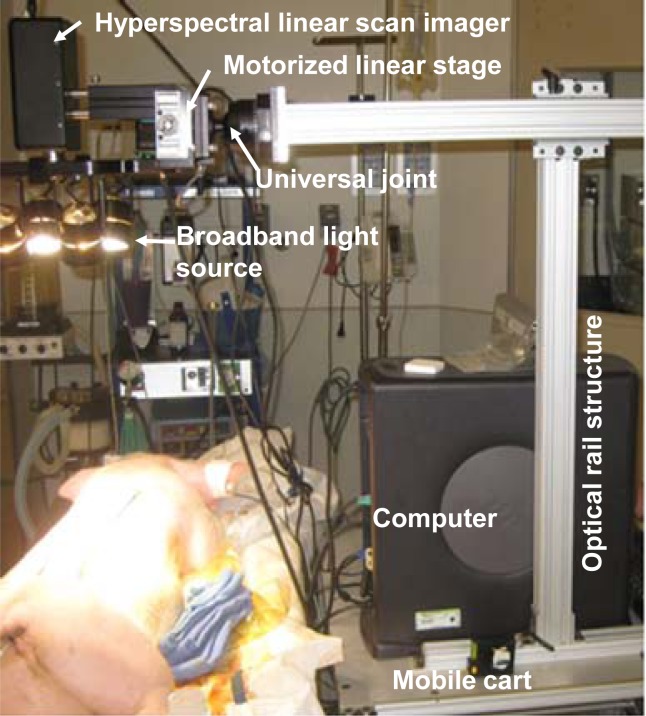
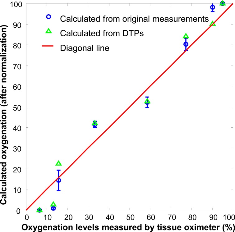
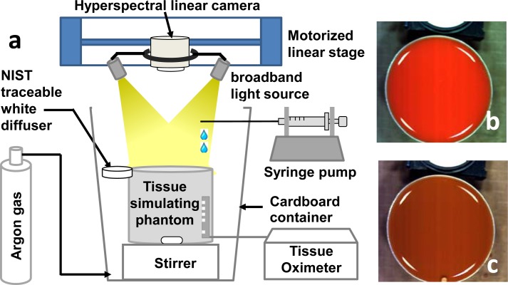
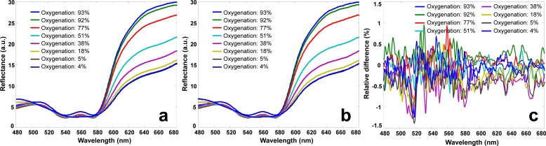
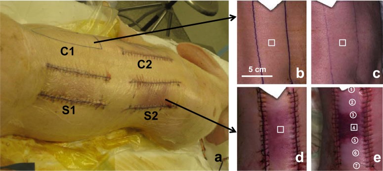
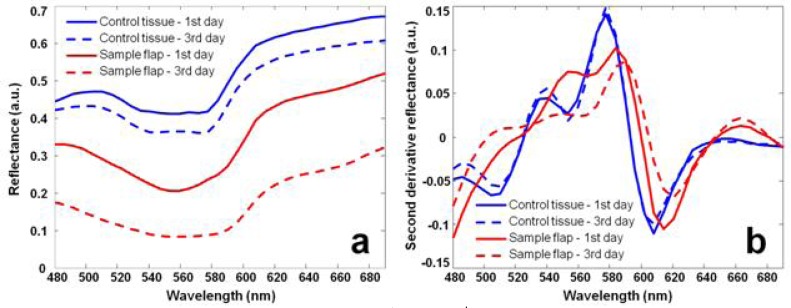
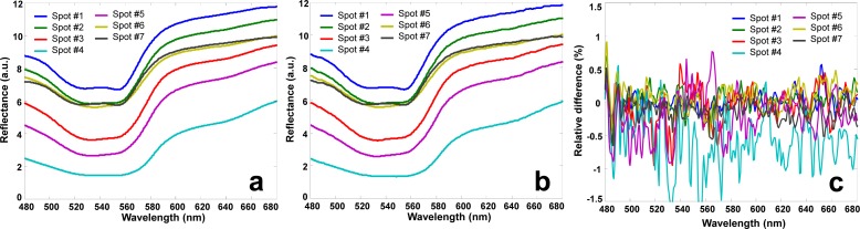
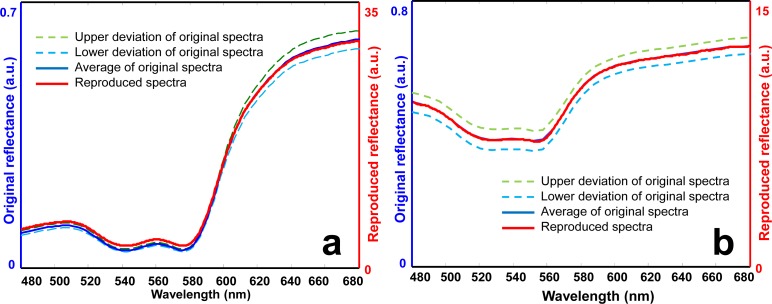
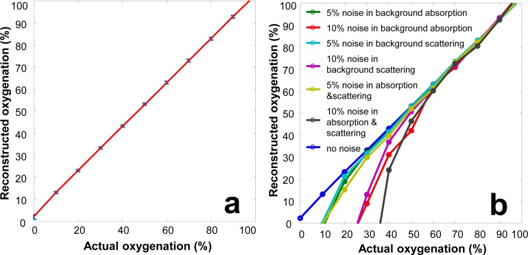
Hyperspectral imaging has the potential to achieve high spatial resolution and high functional sensitivity for non-invasive assessment of tissue oxygenation. However, clinical acceptance of hyperspectral imaging in ischemic wound assessment is hampered by its poor reproducibility, low accuracy, and misinterpreted biology. These limitations are partially caused by the lack of a traceable calibration standard. We proposed a digital tissue phantom (DTP) platform for quantitative calibration and performance evaluation of spectral wound imaging devices. The technical feasibility of such a DTP platform was demonstrated by both in vitro and in vivo experiments. The in vitro DTPs were developed based on a liquid blood phantom model. The in vivo DTPs were developed based on a porcine ischemic skin flap model. The DTPs were projected by a Hyperspectral Image Projector (HIP) with high fidelity. A wide-gap 2nd derivative oxygenation algorithm was developed to reconstruct tissue functional parameters from hyperspectral measurements. In this study, we have demonstrated not only the technical feasibility of using DTPs for quantitative calibration, evaluation, and optimization of spectral imaging devices but also its potential for ischemic wound assessment in clinical practice.
高光谱成像有潜力实现高空间分辨率和高功能灵敏度,用于对组织氧合进行无创评估。然而,高光谱成像在缺血性伤口评估中的临床应用受到其可重复性差、准确性低和生物学解读错误的阻碍。这些局限性部分是由于缺乏可追溯的校准标准所致。我们提出了一个数字组织模型(DTP)平台,用于光谱伤口成像设备的定量校准和性能评估。体外和体内实验均证明了这种DTP平台的技术可行性。体外DTP基于液体血液模型开发。体内DTP基于猪缺血性皮瓣模型开发。DTP由高光谱图像投影仪(HIP)进行高保真投影。开发了一种宽间隙二阶导数氧合算法,用于从高光谱测量中重建组织功能参数。在本研究中,我们不仅证明了使用DTP进行光谱成像设备的定量校准、评估和优化的技术可行性,还证明了其在临床实践中用于缺血性伤口评估的潜力。