Section on Functional Imaging Methods, Laboratory of Brain and Cognition, National Institute of Mental Health, Bldg 10, Rm 1D80, 10 Center Dr MSC 1148, Bethesda, MD 20892-1148, USA.
Neuroimage. 2012 Nov 15;63(3):1712-9. doi: 10.1016/j.neuroimage.2012.06.078. Epub 2012 Jul 14.
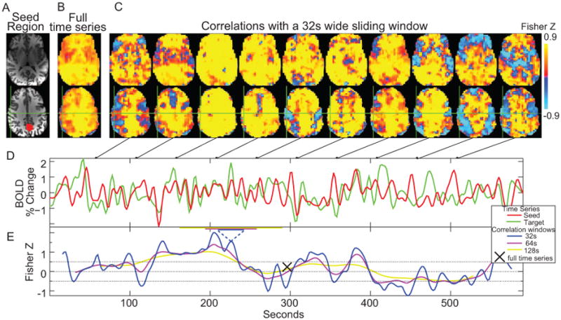
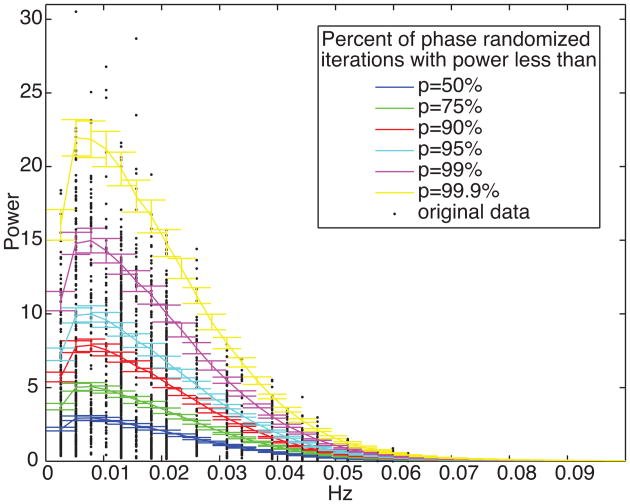
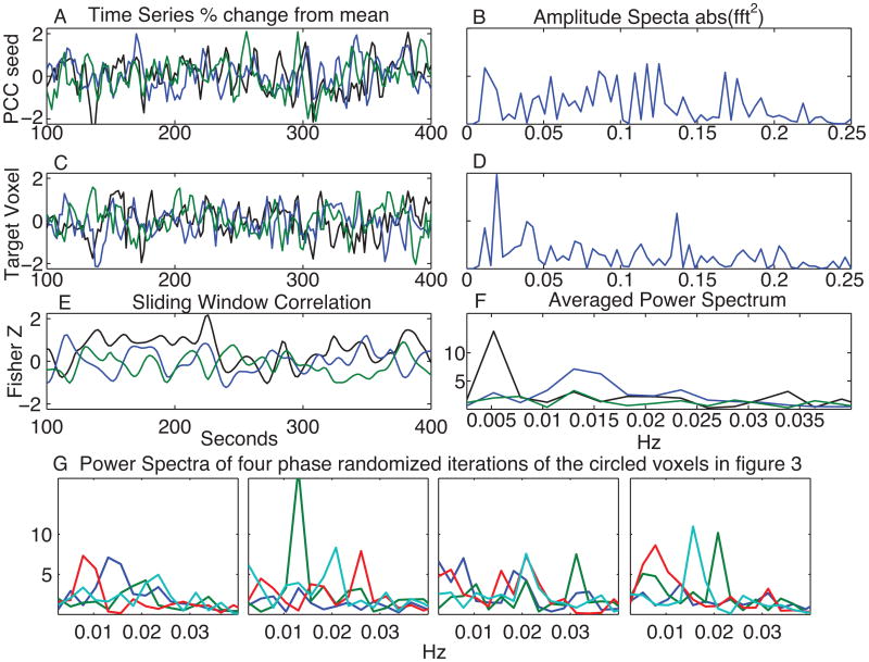
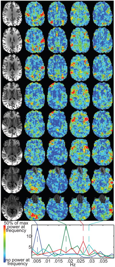
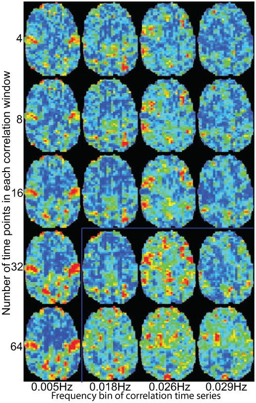
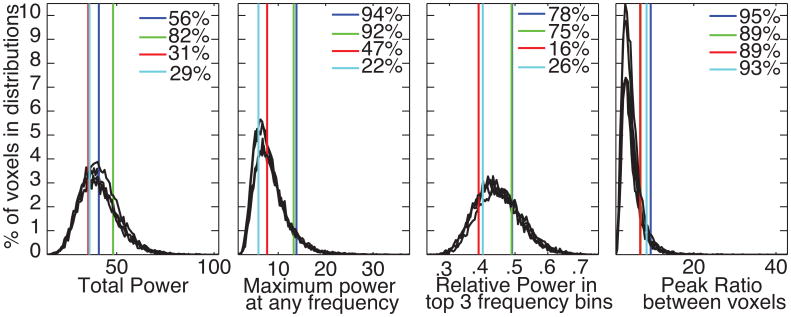
The first two decades of brain research using fMRI have been dominated by studies that measure signal changes in response to a presented task. A rapidly increasing number of studies are showing that consistent activation maps appear by assessment of signal correlations during time periods in which the subjects were not directed to perform any specific task (i.e. "resting state correlations"). Even though neural interactions can happen on much shorter time scales, most "resting state" studies assess these temporal correlations over a period of about 5 to 10 min. Here we investigate how these temporal correlations change on a shorter time scale. We examine changes in brain correlations to the posterior cingulate cortex (PCC) across a 10-minute scan. We show: (1) fMRI correlations fluctuate over time, (2) these fluctuations can be periodic, and (3) correlations between the PCC and other brain regions fluctuate at distinct frequencies. While the precise frequencies of correlation fluctuations vary across subjects and runs, it is still possible to parse brain regions and combinations of brain regions based on fluctuation frequency differences. To evaluate the potential biological significance of these empirical observations, we then use synthetic time series data with identical amplitude spectra, but randomized phase to show that similar effects can still appear even if the timing relationships between voxels are randomized. This implies that observed correlation fluctuations could occur between regions with distinct amplitude spectra, whether or not there are dynamic changes in neural connectivity between such regions. As more studies of brain connectivity dynamics appear, particularly studies using correlation as a key metric, it is vital to better distinguish true neural connectivity dynamics from connectivity fluctuations that are inherently part of this method. Our results also highlight the rich information in the power spectra of fMRI data that can be used to parse brain regions.
使用 fMRI 进行的大脑研究的头二十年主要集中在测量对呈现任务的反应的信号变化的研究上。越来越多的研究表明,在没有指示受试者执行任何特定任务的时间段(即“静息状态相关性”)内,通过评估信号相关性,可以出现一致的激活图。尽管神经相互作用可以在更短的时间尺度上发生,但大多数“静息状态”研究评估这些时间相关性的时间跨度约为 5 到 10 分钟。在这里,我们研究这些时间相关性如何在更短的时间尺度上发生变化。我们检查了在后扣带皮层(PCC)上的脑相关性在 10 分钟扫描过程中的变化。我们表明:(1)fMRI 相关性随时间波动,(2)这些波动可以是周期性的,(3)PCC 与其他脑区之间的相关性以不同的频率波动。虽然相关性波动的精确频率因受试者和运行而异,但仍然可以根据波动频率的差异来解析脑区和脑区的组合。为了评估这些经验观察的潜在生物学意义,我们然后使用具有相同幅度谱但相位随机化的合成时间序列数据来表明,即使在体素之间的时间关系随机化的情况下,仍然可以出现类似的效果。这意味着即使在这些区域之间没有神经连接性的动态变化,观察到的相关性波动也可能发生在具有不同幅度谱的区域之间。随着越来越多的大脑连接动力学研究出现,特别是使用相关性作为关键指标的研究,更好地区分真正的神经连接动力学与固有地属于该方法的连接波动至关重要。我们的结果还突出了 fMRI 数据的功率谱中可以用于解析脑区的丰富信息。