Derix Johanna, Iljina Olga, Schulze-Bonhage Andreas, Aertsen Ad, Ball Tonio
Epilepsy Center, University Medical Center Freiburg Freiburg, Germany.
Front Hum Neurosci. 2012 Sep 5;6:251. doi: 10.3389/fnhum.2012.00251. eCollection 2012.
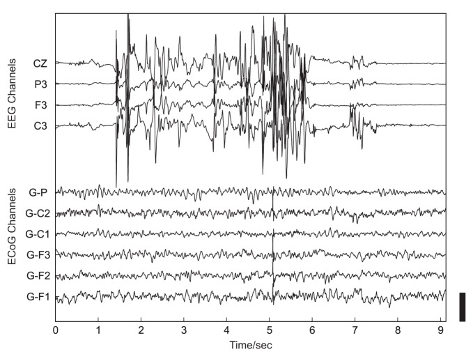
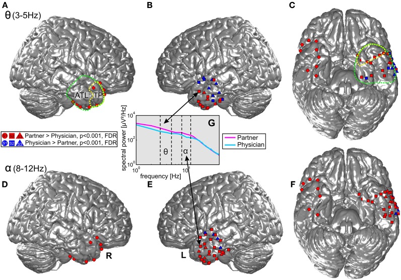
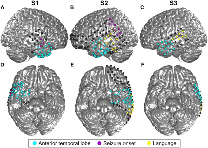
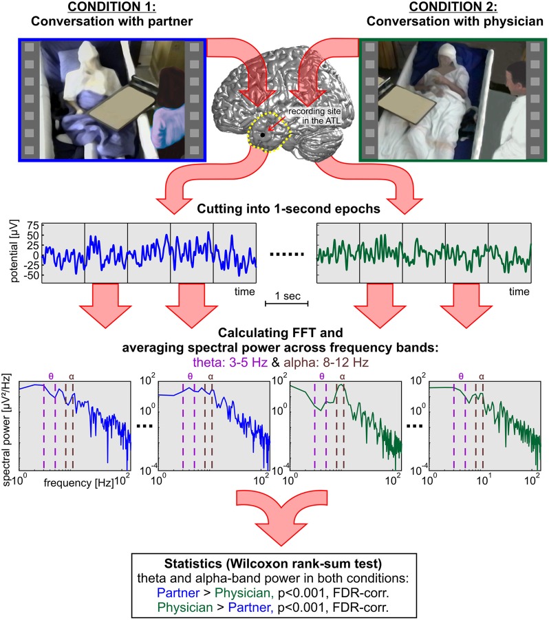
Human brain processes underlying real-life social interaction in everyday situations have been difficult to study and have, until now, remained largely unknown. Here, we investigated whether electrocorticography (ECoG) recorded for pre-neurosurgical diagnostics during the daily hospital life of epilepsy patients could provide a way to elucidate the neural correlates of non-experimental social interaction. We identified time periods in which patients were involved in conversations with either their respective life partners (Condition 1; C1) or attending physicians (Condition 2; C2). These two conditions can be expected to differentially involve subfunctions of social interaction which have been associated with activity in the anterior temporal lobe (ATL), including the temporal pole (TP). Therefore, we specifically focused on ECoG recordings from this brain region and investigated spectral power modulations in the alpha (8-12 Hz) and theta (3-5 Hz) frequency ranges, which have been previously assumed to play an important role in the processing of social interaction. We hypothesized that brain activity in this region might be sensitive to differences in the two interaction situations and tested whether these differences can be detected by single-trial decoding. Condition-specific effects in both theta and alpha bands were observed: the left and right TP exclusively showed increased power in C1 compared to C2, whereas more posterior parts of the ATL exhibited similar (C1 > C2) and also contrary (C2 > C1) effects. Single-trial decoding accuracies for classification of these effects were highly above chance. Our findings demonstrate that it is possible to study the neural correlates of human social interaction in non-experimental conditions. Decoding the identity of the communication partner and adjusting the speech output accordingly may be useful in the emerging field of brain-machine interfacing for restoration of expressive speech.
在日常情境中,人类大脑处理现实生活中社交互动的过程一直难以研究,迄今为止在很大程度上仍不为人知。在此,我们研究了在癫痫患者日常住院生活期间为神经外科手术前诊断记录的皮层脑电图(ECoG)是否能够提供一种方法来阐明非实验性社交互动的神经关联。我们确定了患者与各自生活伴侣(条件1;C1)或主治医生(条件2;C2)进行对话的时间段。可以预期这两种条件会不同程度地涉及社交互动的子功能,这些子功能与颞叶前部(ATL)包括颞极(TP)的活动相关。因此,我们特别关注该脑区的ECoG记录,并研究了先前假定在社交互动处理中起重要作用的α(8 - 12赫兹)和θ(3 - 5赫兹)频率范围内的频谱功率调制。我们假设该区域的大脑活动可能对两种互动情境中的差异敏感,并测试了这些差异是否可以通过单次试验解码检测到。在θ和α波段均观察到特定条件效应:与C2相比,左右TP在C1中仅显示功率增加,而ATL更靠后的部分表现出相似(C1 > C2)以及相反(C2 > C1)的效应。对这些效应进行分类的单次试验解码准确率远高于随机水平。我们的研究结果表明,在非实验条件下研究人类社交互动的神经关联是可能的。解码通信伙伴的身份并相应调整语音输出可能在用于恢复表达性言语的脑机接口新兴领域中有用。