Department of Interventional Therapy, Sun Yat-Sen Memorial Hospital of Sun Yat-Sen University, Guangzhou.
Int J Nanomedicine. 2013;8:3795-804. doi: 10.2147/IJN.S50373. Epub 2013 Oct 2.
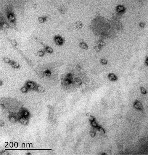
The purpose of this study was to observe the effect and feasibility of hyperthermia and the influence of heat on surrounding organs in a VX2 rabbit liver model exposed to an alternating magnetic field after embolization with ferromagnetic nanoparticles.
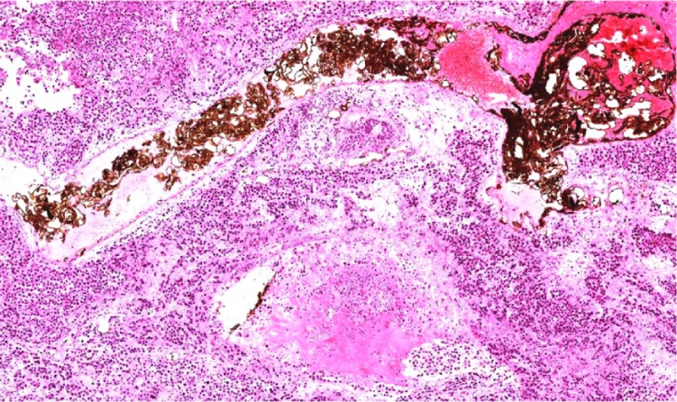
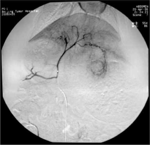
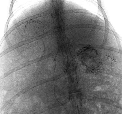
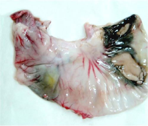
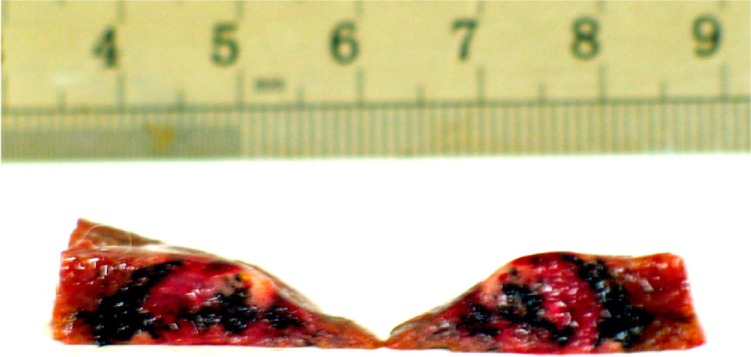
Forty rabbits containing implanted hepatic VX2 carcinomas were divided into four groups, each containing ten rabbits. Fourteen days after tumor transplantation, we opened the abdomen to observe the size and shape of the tumor. A transfemoral retrograde approach was then used for hepatic arterial catheterization in groups B, C, and D to perform angiography and embolization. The next day, three rabbits in group B and all rabbits in group D were exposed to an alternating magnetic field, and the temperature was recorded simultaneously in the center of the tumor, at the edge of the tumor, and in the normal liver parenchyma. On day 28, all animals was euthanized to observe changes in the implanted liver tumor and the condition of the abdomen. A pathologic examination was also done.
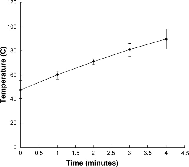
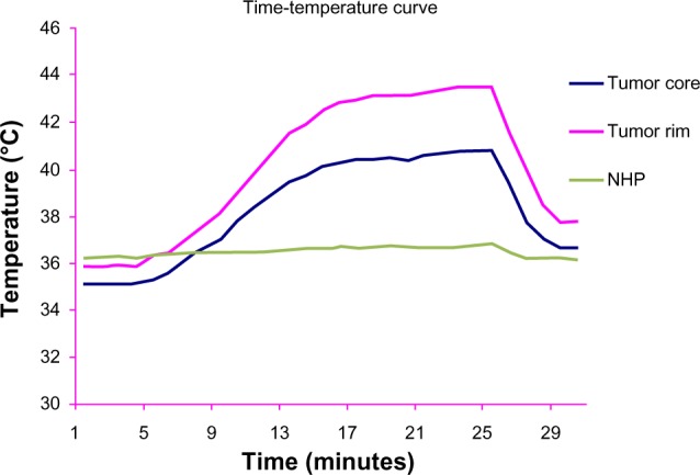
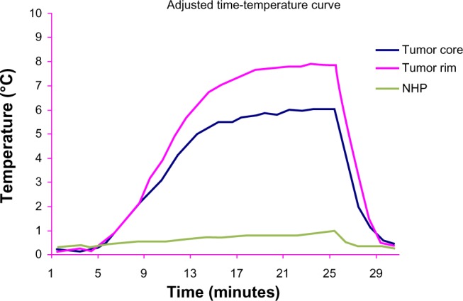
Before surgery, there was no significant difference in tumor volume between the four groups. Three different temperature points (cen ter of the tumor, edge of the tumor, and in the normal liver parenchyma) of group B under an alternating magnetic field were 37.2°C ± 1.1°C, 36.8°C ± 1.2°C, and 36.9°C ± 2.1°C, none of which were significantly different from pretreatment values. Three points basal temperature in group D showed no significant difference (F = 1.038, P = 0.413). Seven to 26 minutes after hyperthermia, the temperature at the center of the tumor and at the edge of the tumor in group D was significantly different from the corresponding points in group B and from normal liver tissue in group D (F(B-D center) = 5.431, P(B-D center) = 0.041, F(B-D edge) = 9.744, P(B-D edge) = 0.011; F(D) = 8.379, P(D) = 0.002). The highest temperature recorded at the rim of the tumor was 46°C in group D. Fourteen days later, the tumor volume in the four groups was group A 31.4 ± 20.6 cm(3), group B 26.7 ± 18.2 cm(3), group C 28.7 ± 9.1 cm(3), and group D 25.8 ± 13.9 cm(3), with no significant difference found between the groups (F = 0.218, P = 0.883). The increase in tumor volume was greatest in group A and least in group D, while that in groups B and D was similar.
It is feasible to treat a VX2 tumor in an alternating magnetic field after embolization with magnetic nanoparticles without a significant effect on the surrounding normal liver parenchyma.
本研究旨在观察在栓塞铁磁纳米颗粒后,交变磁场对兔 VX2 肝癌模型中热疗的效果和可行性,以及对周围器官的影响。
40 只荷有植入性肝 VX2 癌的兔被分为 4 组,每组 10 只。肿瘤移植后 14 天,我们打开腹部观察肿瘤的大小和形状。然后,通过经股逆行肝动脉插管,对 B、C、D 组进行血管造影和栓塞。第二天,B 组的 3 只兔子和 D 组的所有兔子都暴露在交变磁场中,同时记录肿瘤中心、肿瘤边缘和正常肝组织的温度。第 28 天,所有动物被安乐死,观察植入性肝肿瘤和腹部情况的变化。还进行了病理检查。
手术前,4 组肿瘤体积无明显差异。在交变磁场下,B 组的 3 个不同温度点(肿瘤中心、肿瘤边缘和正常肝组织)分别为 37.2°C ± 1.1°C、36.8°C ± 1.2°C 和 36.9°C ± 2.1°C,均与术前值无显著差异。D 组的 3 个基础温度点无显著差异(F = 1.038,P = 0.413)。热疗后 7 至 26 分钟,D 组肿瘤中心和边缘的温度与 B 组相应点和 D 组正常肝组织的温度有显著差异(F(B-D 中心)= 5.431,P(B-D 中心)= 0.041,F(B-D 边缘)= 9.744,P(B-D 边缘)= 0.011;F(D)= 8.379,P(D)= 0.002)。D 组肿瘤边缘记录的最高温度为 46°C。14 天后,4 组肿瘤体积分别为 A 组 31.4 ± 20.6 cm(3)、B 组 26.7 ± 18.2 cm(3)、C 组 28.7 ± 9.1 cm(3)和 D 组 25.8 ± 13.9 cm(3),组间无显著差异(F = 0.218,P = 0.883)。肿瘤体积增加最大的是 A 组,最小的是 D 组,而 B 组和 D 组的增加相似。
在栓塞铁磁纳米颗粒后,用交变磁场治疗 VX2 肿瘤是可行的,对周围正常肝组织没有明显影响。