Department of Radiology and Biomedical Imaging, University of California San Francisco, San Francisco, California, United States of America.
PLoS One. 2013 Dec 4;8(12):e81653. doi: 10.1371/journal.pone.0081653. eCollection 2013.
Clinical scores of mammographic breast density are highly subjective. Automated technologies for mammography exist to quantify breast density objectively, but the technique that most accurately measures the quantity of breast fibroglandular tissue is not known.
To compare the agreement of three automated mammographic techniques for measuring volumetric breast density with a quantitative volumetric MRI-based technique in a screening population.
Women were selected from the UCSF Medical Center screening population that had received both a screening MRI and digital mammogram within one year of each other, had Breast Imaging Reporting and Data System (BI-RADS) assessments of normal or benign finding, and no history of breast cancer or surgery. Agreement was assessed of three mammographic techniques (Single-energy X-ray Absorptiometry [SXA], Quantra, and Volpara) with MRI for percent fibroglandular tissue volume, absolute fibroglandular tissue volume, and total breast volume.
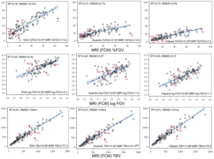
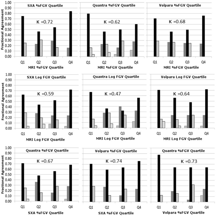
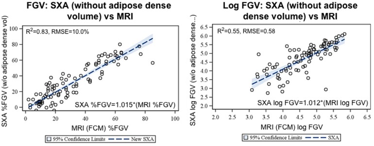
Among 99 women, the automated mammographic density techniques were correlated with MRI measures with R(2) values ranging from 0.40 (log fibroglandular volume) to 0.91 (total breast volume). Substantial agreement measured by kappa statistic was found between all percent fibroglandular tissue measures (0.72 to 0.63), but only moderate agreement for log fibroglandular volumes. The kappa statistics for all percent density measures were highest in the comparisons of the SXA and MRI results. The largest error source between MRI and the mammography techniques was found to be differences in measures of total breast volume.
Automated volumetric fibroglandular tissue measures from screening digital mammograms were in substantial agreement with MRI and if associated with breast cancer could be used in clinical practice to enhance risk assessment and prevention.
乳腺 X 线摄影的临床评分具有高度主观性。存在用于客观量化乳腺密度的自动化技术,但尚不清楚哪种技术最能准确测量乳腺纤维腺体组织的数量。
在筛查人群中,比较三种自动乳腺 X 线摄影技术测量体积乳腺密度与基于定量容积磁共振成像技术的一致性。
从 UCSF 医疗中心的筛查人群中选择在彼此的一年之内接受过筛查性 MRI 和数字乳腺 X 线摄影检查、BI-RADS 评估为正常或良性发现且无乳腺癌或手术史的女性。评估了三种乳腺 X 线摄影技术(单能 X 射线吸收法[SXA]、Quantra 和 Volpara)与 MRI 对纤维腺体组织体积百分比、绝对纤维腺体组织体积和总乳房体积的一致性。
在 99 名女性中,自动乳腺 X 线摄影密度技术与 MRI 测量值具有 R(2)值,范围从 0.40(对数纤维腺体体积)到 0.91(总乳房体积)。通过kappa 统计量发现,所有纤维腺体组织百分比测量值之间均存在显著一致性(0.72 至 0.63),但对数纤维腺体体积的一致性仅为中度。SXA 和 MRI 结果比较时,所有百分比密度测量值的kappa 统计量最高。在 MRI 和乳腺 X 线摄影技术之间,最大的误差源是总乳房体积测量值的差异。
筛查性数字乳腺 X 线摄影的自动容积纤维腺体组织测量值与 MRI 具有实质性一致性,如果与乳腺癌相关,则可用于临床实践,以增强风险评估和预防。