• 文献检索
  • 文档翻译
  • 深度研究
  • 学术资讯
  • Suppr Zotero 插件Zotero 插件
  • 邀请有礼
  • 套餐&价格
  • 历史记录
应用&插件
Suppr Zotero 插件Zotero 插件浏览器插件Mac 客户端Windows 客户端微信小程序
定价
高级版会员购买积分包购买API积分包
服务
文献检索文档翻译深度研究API 文档MCP 服务
关于我们
关于 Suppr公司介绍联系我们用户协议隐私条款
关注我们

Suppr 超能文献

核心技术专利:CN118964589B侵权必究
粤ICP备2023148730 号-1Suppr @ 2026

文献检索

告别复杂PubMed语法,用中文像聊天一样搜索,搜遍4000万医学文献。AI智能推荐,让科研检索更轻松。

立即免费搜索

文件翻译

保留排版,准确专业,支持PDF/Word/PPT等文件格式,支持 12+语言互译。

免费翻译文档

深度研究

AI帮你快速写综述,25分钟生成高质量综述,智能提取关键信息,辅助科研写作。

立即免费体验

T1加权和T2加权磁共振图像的纹理分析以及使用概率神经网络鉴别儿童后颅窝肿瘤

Texture analysis of T1 - and T2 -weighted MR images and use of probabilistic neural network to discriminate posterior fossa tumours in children.

作者信息

Orphanidou-Vlachou Eleni, Vlachos Nikolaos, Davies Nigel P, Arvanitis Theodoros N, Grundy Richard G, Peet Andrew C

机构信息

Birmingham Children's Hospital NHS Foundation Trust, Birmingham, UK; School of Cancer Sciences, College of Medical and Dental Sciences, University of Birmingham, Edgbaston, Birmingham, UK.

出版信息

NMR Biomed. 2014 Jun;27(6):632-9. doi: 10.1002/nbm.3099. Epub 2014 Apr 13.

DOI:10.1002/nbm.3099
PMID:24729528
原文链接:https://pmc.ncbi.nlm.nih.gov/articles/PMC4529665/
Abstract

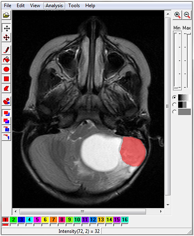
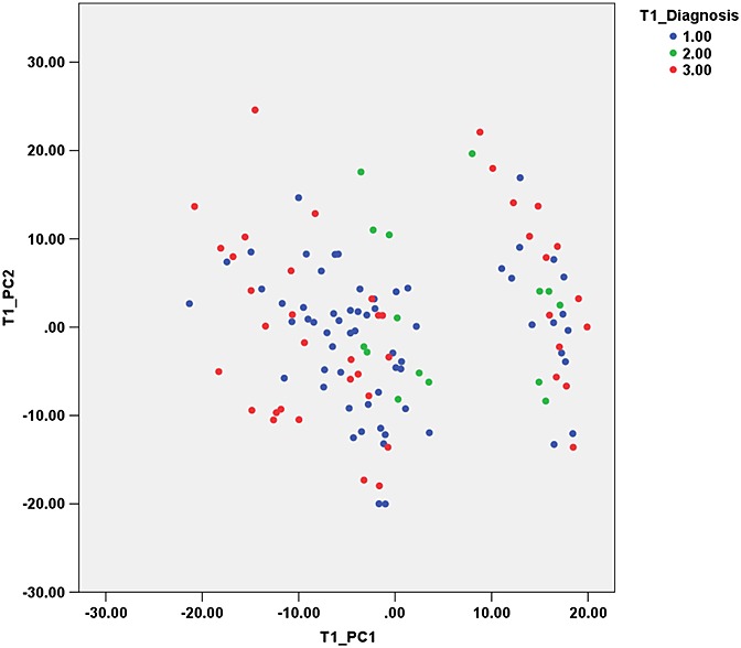
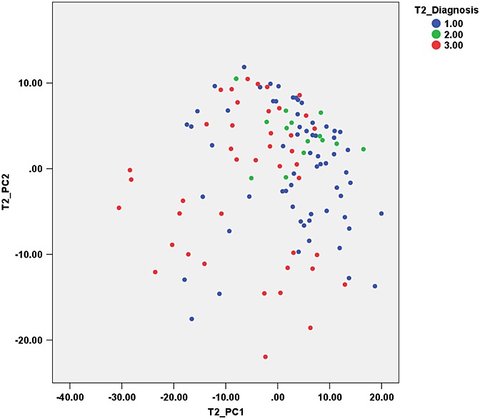
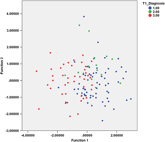
Brain tumours are the most common solid tumours in children, representing 20% of all cancers. The most frequent posterior fossa tumours are medulloblastomas, pilocytic astrocytomas and ependymomas. Texture analysis (TA) of MR images can be used to support the diagnosis of these tumours by providing additional quantitative information. MaZda software was used to perform TA on T1 - and T2 -weighted images of children with pilocytic astrocytomas, medulloblastomas and ependymomas of the posterior fossa, who had MRI at Birmingham Children's Hospital prior to treatment. The region of interest was selected on three slices per patient in Image J, using thresholding and manual outlining. TA produced 279 features, which were reduced using principal component analysis (PCA). The principal components (PCs) explaining 95% of the variance were used in a linear discriminant analysis (LDA) and a probabilistic neural network (PNN) to classify the cases, using DTREG statistics software. PCA of texture features from both T1 - and T2 -weighted images yielded 13 PCs to explain >95% of the variance. The PNN classifier for T1 -weighted images achieved 100% accuracy on training the data and 90% on leave-one-out cross-validation (LOOCV); for T2 -weighted images, the accuracy was 100% on training the data and 93.3% on LOOCV. A PNN classifier with T1 and T2 PCs achieved 100% accuracy on training the data and 85.8% on LOOCV. LDA classification accuracies were noticeably poorer. The features found to hold the highest discriminating potential were all co-occurrence matrix derived, where adjacent pixels had highly correlated intensities. This study shows that TA can be performed on standard T1 - and T2 -weighted images of childhood posterior fossa tumours using readily available software to provide high diagnostic accuracy. Discriminatory features do not correspond to those used in the clinical interpretation of the images and therefore provide novel tumour information.

摘要

脑肿瘤是儿童中最常见的实体肿瘤,占所有癌症的20%。最常见的后颅窝肿瘤是髓母细胞瘤、毛细胞型星形细胞瘤和室管膜瘤。磁共振成像(MR)图像的纹理分析(TA)可通过提供额外的定量信息来辅助这些肿瘤的诊断。使用MaZda软件对在伯明翰儿童医院治疗前进行过MRI检查的患有后颅窝毛细胞型星形细胞瘤、髓母细胞瘤和室管膜瘤的儿童的T1加权和T2加权图像进行TA。在Image J中,通过阈值处理和手动勾勒轮廓,为每位患者在三个切片上选择感兴趣区域。TA产生了279个特征,使用主成分分析(PCA)对这些特征进行了降维。使用DTREG统计软件,将解释95%方差的主成分(PC)用于线性判别分析(LDA)和概率神经网络(PNN)对病例进行分类。来自T1加权和T2加权图像的纹理特征的PCA产生了13个PC来解释>95%的方差。T1加权图像的PNN分类器在训练数据时准确率达到100%,在留一法交叉验证(LOOCV)时为90%;对于T2加权图像,训练数据时准确率为100%,LOOCV时为93.3%。结合T1和T2主成分的PNN分类器在训练数据时准确率达到100%,LOOCV时为85.8%。LDA分类准确率明显较低。发现具有最高鉴别潜力的特征均来自共生矩阵,其中相邻像素具有高度相关的强度。本研究表明,使用现成的软件可以对儿童后颅窝肿瘤的标准T1加权和T2加权图像进行TA,以提供高诊断准确性。鉴别特征与图像临床解读中使用的特征不同,因此提供了新的肿瘤信息。

https://cdn.ncbi.nlm.nih.gov/pmc/blobs/7942/4529665/fb04beb7a380/nbm0027-0632-f5.jpg
https://cdn.ncbi.nlm.nih.gov/pmc/blobs/7942/4529665/853530aa9136/nbm0027-0632-f1.jpg
https://cdn.ncbi.nlm.nih.gov/pmc/blobs/7942/4529665/e66fe09fac3d/nbm0027-0632-f2.jpg
https://cdn.ncbi.nlm.nih.gov/pmc/blobs/7942/4529665/4fadfabbb30a/nbm0027-0632-f3.jpg
https://cdn.ncbi.nlm.nih.gov/pmc/blobs/7942/4529665/35123052222b/nbm0027-0632-f4.jpg
https://cdn.ncbi.nlm.nih.gov/pmc/blobs/7942/4529665/fb04beb7a380/nbm0027-0632-f5.jpg

相似文献

1
Texture analysis of T1 - and T2 -weighted MR images and use of probabilistic neural network to discriminate posterior fossa tumours in children.T1加权和T2加权磁共振图像的纹理分析以及使用概率神经网络鉴别儿童后颅窝肿瘤
NMR Biomed. 2014 Jun;27(6):632-9. doi: 10.1002/nbm.3099. Epub 2014 Apr 13.
2
Three-dimensional textural features of conventional MRI improve diagnostic classification of childhood brain tumours.传统MRI的三维纹理特征可改善儿童脑肿瘤的诊断分类。
NMR Biomed. 2015 Sep;28(9):1174-84. doi: 10.1002/nbm.3353. Epub 2015 Aug 9.
3
Metrics and textural features of MRI diffusion to improve classification of pediatric posterior fossa tumors.磁共振成像扩散的指标和纹理特征以改善小儿后颅窝肿瘤的分类
AJNR Am J Neuroradiol. 2014 May;35(5):1009-15. doi: 10.3174/ajnr.A3784. Epub 2013 Dec 5.
4
Magnetic Resonance Imaging Features of Common Posterior Fossa Brain Tumors in Children: A Preliminary Vietnamese Study.儿童常见后颅窝脑肿瘤的磁共振成像特征:一项越南初步研究。
Open Access Maced J Med Sci. 2019 Aug 14;7(15):2413-2418. doi: 10.3889/oamjms.2019.635. eCollection 2019 Aug 15.
5
3D Texture Analysis of Heterogeneous MRI Data for Diagnostic Classification of Childhood Brain Tumours.用于儿童脑肿瘤诊断分类的异质性MRI数据的3D纹理分析
Stud Health Technol Inform. 2015;213:19-22.
6
Convolutional neural network-based magnetic resonance image differentiation of filum terminale ependymomas from schwannomas.基于卷积神经网络的马尾神经室管膜瘤与神经鞘瘤的磁共振图像鉴别。
BMC Cancer. 2024 Mar 19;24(1):350. doi: 10.1186/s12885-024-12023-0.
7
MR signal of the solid portion of pilocytic astrocytoma on T2-weighted images: is it useful for differentiation from medulloblastoma?毛细胞型星形细胞瘤实性部分在T2加权图像上的磁共振信号:对与髓母细胞瘤鉴别诊断有用吗?
Neuroradiology. 2006 Apr;48(4):233-7. doi: 10.1007/s00234-006-0048-5. Epub 2006 Mar 21.
8
Robust texture features for response monitoring of glioblastoma multiforme on T1-weighted and T2-FLAIR MR images: a preliminary investigation in terms of identification and segmentation.用于 T1 加权和 T2-FLAIR MR 图像胶质母细胞瘤反应监测的稳健纹理特征:在识别和分割方面的初步研究。
Med Phys. 2010 Apr;37(4):1722-36. doi: 10.1118/1.3357289.
9
MRI findings of olivary degeneration after surgery for posterior fossa tumours in children: incidence, time course and correlation with tumour grading.儿童后颅窝肿瘤手术后橄榄核变性的 MRI 表现:发生率、时间过程及与肿瘤分级的相关性。
Radiol Med. 2015 May;120(5):474-82. doi: 10.1007/s11547-014-0477-x. Epub 2015 Jan 9.
10
Diagnostic accuracy of texture analysis applied to T- and T-Relaxation maps for liver fibrosis classification via machine-learning algorithms with liver histology as reference standard.以肝脏组织学为参考标准,通过机器学习算法,应用于T1和T2弛豫图的纹理分析对肝纤维化分类的诊断准确性。
Eur J Radiol. 2025 Feb;183:111887. doi: 10.1016/j.ejrad.2024.111887. Epub 2024 Dec 20.

引用本文的文献

1
Diagnostic Performance of Neural Network-Based Artificial Intelligence in the Detection and Classification of Pediatric Astrocytoma: A Systematic Review.基于神经网络的人工智能在小儿星形细胞瘤检测与分类中的诊断性能:一项系统评价
Cureus. 2025 May 5;17(5):e83543. doi: 10.7759/cureus.83543. eCollection 2025 May.
2
The Role of Machine Learning Approaches in Pediatric Oncology: A Systematic Review.机器学习方法在儿科肿瘤学中的作用:一项系统综述。
Cureus. 2025 Jan 16;17(1):e77524. doi: 10.7759/cureus.77524. eCollection 2025 Jan.
3
Knowledge discovery from database: MRI radiomic features to assess recurrence risk in high-grade meningiomas.

本文引用的文献

1
Accurate classification of childhood brain tumours by in vivo ¹H MRS - a multi-centre study.¹H MRS 体内技术对儿童脑肿瘤的准确分类——一项多中心研究。
Eur J Cancer. 2013 Feb;49(3):658-67. doi: 10.1016/j.ejca.2012.09.003. Epub 2012 Oct 2.
2
Discrimination of paediatric brain tumours using apparent diffusion coefficient histograms.基于表观扩散系数直方图鉴别小儿脑肿瘤。
Eur Radiol. 2012 Feb;22(2):447-57. doi: 10.1007/s00330-011-2255-7. Epub 2011 Sep 15.
3
Value and limitations of diffusion-weighted imaging in grading and diagnosis of pediatric posterior fossa tumors.
从数据库中发现知识:用于评估高级别脑膜瘤复发风险的MRI影像组学特征
BMC Med Imaging. 2025 Jan 9;25(1):14. doi: 10.1186/s12880-024-01483-2.
4
Application of radiomics for diagnosis, subtyping, and prognostication of medulloblastomas: a systematic review.基于放射组学的脑桥小脑角脑膜瘤诊断、分型及预后评估的系统评价
Neurosurg Rev. 2024 Oct 29;47(1):827. doi: 10.1007/s10143-024-03060-1.
5
Metabolite profiles of medulloblastoma for rapid and non-invasive detection of molecular disease groups.用于快速无创检测分子疾病组的髓母细胞瘤代谢物图谱。
EBioMedicine. 2024 Feb;100:104958. doi: 10.1016/j.ebiom.2023.104958. Epub 2024 Jan 6.
6
Deciphering Machine Learning Decisions to Distinguish between Posterior Fossa Tumor Types Using MRI Features: What Do the Data Tell Us?利用MRI特征解读机器学习决策以区分后颅窝肿瘤类型:数据告诉了我们什么?
Cancers (Basel). 2023 Aug 8;15(16):4015. doi: 10.3390/cancers15164015.
7
Applications of artificial intelligence in magnetic resonance imaging of primary pediatric cancers: a scoping review and CLAIM score assessment.人工智能在原发性儿科癌症磁共振成像中的应用:范围综述和 CLAIM 评分评估。
Jpn J Radiol. 2023 Oct;41(10):1127-1147. doi: 10.1007/s11604-023-01437-8. Epub 2023 Jul 3.
8
Machine Learning in the Classification of Pediatric Posterior Fossa Tumors: A Systematic Review.机器学习在小儿后颅窝肿瘤分类中的应用:一项系统综述。
Cancers (Basel). 2022 Nov 15;14(22):5608. doi: 10.3390/cancers14225608.
9
The role of artificial intelligence in paediatric neuroradiology.人工智能在儿科神经放射学中的作用。
Pediatr Radiol. 2022 Oct;52(11):2159-2172. doi: 10.1007/s00247-022-05322-w. Epub 2022 Mar 26.
10
Beyond Glioma: The Utility of Radiomic Analysis for Non-Glial Intracranial Tumors.超越胶质瘤:放射组学分析在非胶质细胞性颅内肿瘤中的应用
Cancers (Basel). 2022 Feb 7;14(3):836. doi: 10.3390/cancers14030836.
弥散加权成像在儿童后颅窝肿瘤分级和诊断中的价值及局限性。
AJNR Am J Neuroradiol. 2010 Oct;31(9):1613-6. doi: 10.3174/ajnr.A2155. Epub 2010 Jun 10.
4
Texture analysis: a review of neurologic MR imaging applications.纹理分析:神经磁共振成像应用综述。
AJNR Am J Neuroradiol. 2010 May;31(5):809-16. doi: 10.3174/ajnr.A2061. Epub 2010 Apr 15.
5
Magnetic resonance spectroscopy in pediatric neuroradiology: clinical and research applications.磁共振波谱在儿科神经放射学中的临床与研究应用
Pediatr Radiol. 2010 Jan;40(1):3-30. doi: 10.1007/s00247-009-1450-z. Epub 2009 Nov 24.
6
Comparison among probabilistic neural network, support vector machine and logistic regression for evaluating the effect of subthalamic stimulation in Parkinson disease on ground reaction force during gait.概率神经网络、支持向量机和逻辑回归在评估帕金森病患者丘脑底核刺激对步态中地面反力影响的比较。
J Biomech. 2010 Mar 3;43(4):720-6. doi: 10.1016/j.jbiomech.2009.10.018. Epub 2009 Nov 14.
7
Pediatric low-grade gliomas.儿童低级别胶质瘤
J Child Neurol. 2009 Nov;24(11):1397-408. doi: 10.1177/0883073809342005.
8
Neuroimaging of pediatric brain tumors: from basic to advanced magnetic resonance imaging (MRI).小儿脑肿瘤的神经影像学:从基础到高级磁共振成像(MRI)
J Child Neurol. 2009 Nov;24(11):1343-65. doi: 10.1177/0883073809342129.
9
An analysis of image texture, tumor location, and MGMT promoter methylation in glioblastoma using magnetic resonance imaging.基于磁共振成像的脑胶质母细胞瘤图像纹理、肿瘤位置和 MGMT 启动子甲基化分析。
Neuroimage. 2010 Jan 15;49(2):1398-405. doi: 10.1016/j.neuroimage.2009.09.049. Epub 2009 Sep 28.
10
High resolution magic angle spinning 1H NMR of childhood brain and nervous system tumours.儿童脑和神经系统肿瘤的高分辨率魔角旋转1H核磁共振
Mol Cancer. 2009 Feb 10;8:6. doi: 10.1186/1476-4598-8-6.