Hectors Stefanie J C G, Jacobs Igor, Strijkers Gustav J, Nicolay Klaas
Biomedical NMR, Department of Biomedical Engineering, Eindhoven University of Technology, Eindhoven, The Netherlands; Center for Imaging Research and Education (CIRE), Eindhoven, The Netherlands.
Biomedical NMR, Department of Biomedical Engineering, Eindhoven University of Technology, Eindhoven, The Netherlands; Center for Imaging Research and Education (CIRE), Eindhoven, The Netherlands; Biomedical Engineering and Physics, Academic Medical Center, University of Amsterdam, Amsterdam, The Netherlands.
PLoS One. 2014 Jun 13;9(6):e99936. doi: 10.1371/journal.pone.0099936. eCollection 2014.
In this study endogenous magnetic resonance imaging (MRI) biomarkers for accurate segmentation of High Intensity Focused Ultrasound (HIFU)-treated tumor tissue and residual or recurring non-treated tumor tissue were identified.
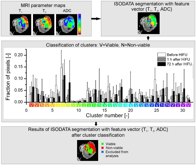
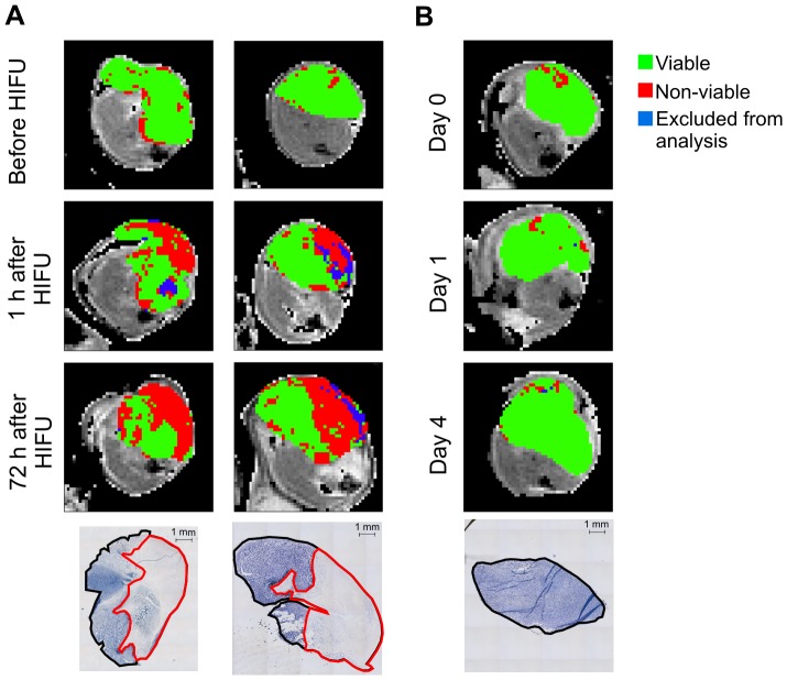
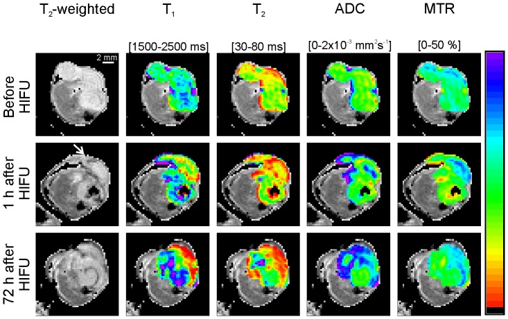
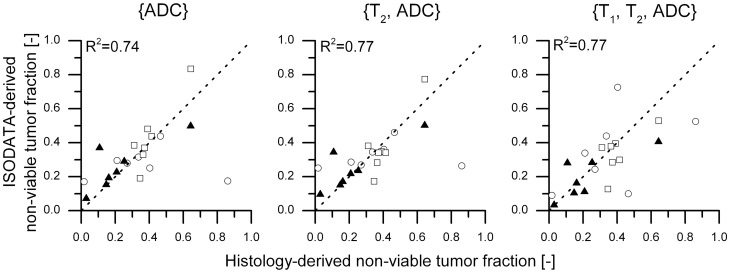
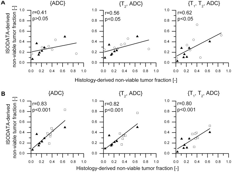
Multiparametric MRI, consisting of quantitative T1, T2, Apparent Diffusion Coefficient (ADC) and Magnetization Transfer Ratio (MTR) mapping, was performed in tumor-bearing mice before (n = 14), 1 h after (n = 14) and 72 h (n = 7) after HIFU treatment. A non-treated control group was included (n = 7). Cluster analysis using the Iterative Self Organizing Data Analysis (ISODATA) technique was performed on subsets of MRI parameters (feature vectors). The clusters resulting from the ISODATA segmentation were divided into a viable and non-viable class based on the fraction of pixels assigned to the clusters at the different experimental time points. ISODATA-derived non-viable tumor fractions were quantitatively compared to histology-derived non-viable tumor volume fractions.
The highest agreement between the ISODATA-derived and histology-derived non-viable tumor fractions was observed for feature vector {T1, T2, ADC}. R1 (1/T1), R2 (1/T2), ADC and MTR each were significantly increased in the ISODATA-defined non-viable tumor tissue at 1 h after HIFU treatment compared to viable, non-treated tumor tissue. R1, ADC and MTR were also significantly increased at 72 h after HIFU.
This study demonstrates that non-viable, HIFU-treated tumor tissue can be distinguished from viable, non-treated tumor tissue using multiparametric MRI analysis. Clinical application of the presented methodology may allow for automated, accurate and objective evaluation of HIFU treatment.
本研究旨在识别用于准确分割高强度聚焦超声(HIFU)治疗的肿瘤组织以及残留或复发性未治疗肿瘤组织的内源性磁共振成像(MRI)生物标志物。
对荷瘤小鼠在HIFU治疗前(n = 14)、治疗后1小时(n = 14)和治疗后72小时(n = 7)进行多参数MRI检查,包括定量T1、T2、表观扩散系数(ADC)和磁化传递率(MTR)成像。纳入一个未治疗的对照组(n = 7)。使用迭代自组织数据分析(ISODATA)技术对MRI参数子集(特征向量)进行聚类分析。根据在不同实验时间点分配给聚类的像素比例,将ISODATA分割产生的聚类分为存活和非存活类别。将ISODATA得出的非存活肿瘤分数与组织学得出的非存活肿瘤体积分数进行定量比较。
对于特征向量{T1, T2, ADC},观察到ISODATA得出的和组织学得出的非存活肿瘤分数之间的一致性最高。与存活的未治疗肿瘤组织相比,在HIFU治疗后1小时,ISODATA定义的非存活肿瘤组织中的R1(1/T1)、R2(1/T2)、ADC和MTR均显著增加。在HIFU治疗后72小时,R1、ADC和MTR也显著增加。
本研究表明,使用多参数MRI分析可以区分HIFU治疗的非存活肿瘤组织和未治疗的存活肿瘤组织。所提出方法的临床应用可能允许对HIFU治疗进行自动化、准确和客观的评估。