Lee Nayeon, Park Jae Woo, Kim Hyung Joon, Yeon Ju Hun, Kwon Jihye, Ko Jung Jae, Oh Seung-Hun, Kim Hyun Sook, Kim Aeri, Han Baek Soo, Lee Sang Chul, Jeon Noo Li, Song Jihwan
CHA Stem Cell Institute, CHA University, Seoul 135-081, Korea.
Mol Cells. 2014 Jun;37(6):497-502. doi: 10.14348/molcells.2014.0137. Epub 2014 Jun 18.
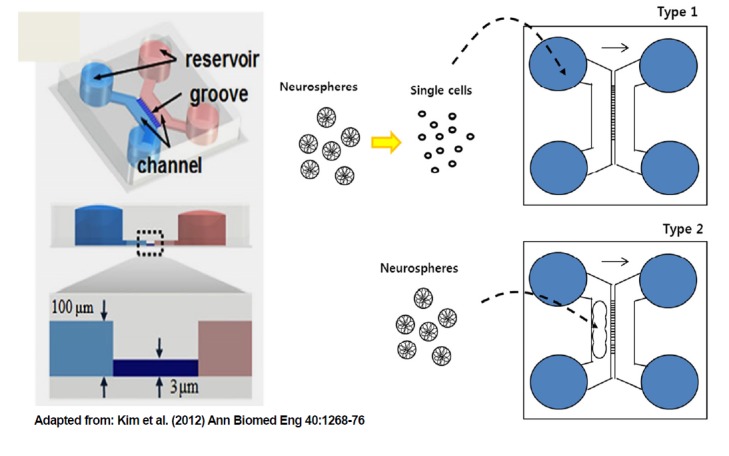
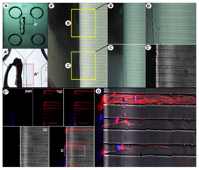
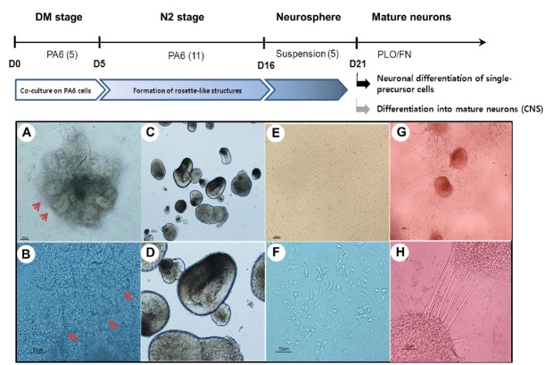
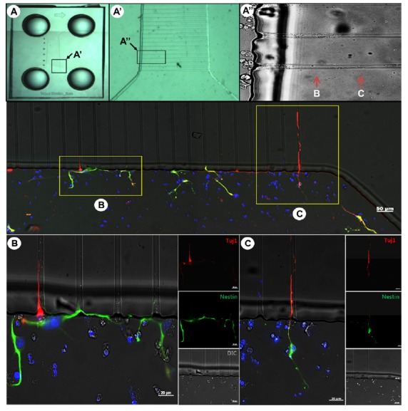
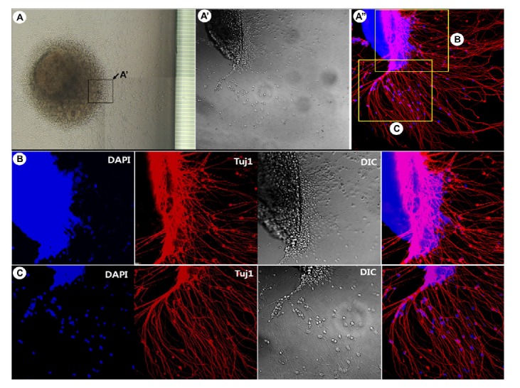
Microfluidics can provide unique experimental tools to visualize the development of neural structures within a microscale device, which is followed by guidance of neurite growth in the axonal isolation compartment. We utilized microfluidics technology to monitor the differentiation and migration of neural cells derived from human embryonic stem cells (hESCs). We co-cultured hESCs with PA6 stromal cells, and isolated neural rosette-like structures, which subsequently formed neurospheres in suspension culture. Tuj1-positive neural cells, but not nestin-positive neural precursor cells (NPCs), were able to enter the microfluidics grooves (microchannels), suggesting that neural cell-migratory capacity was dependent upon neuronal differentiation stage. We also showed that bundles of axons formed and extended into the microchannels. Taken together, these results demonstrated that microfluidics technology can provide useful tools to study neurite outgrowth and axon guidance of neural cells, which are derived from human embryonic stem cells.
微流控技术可以提供独特的实验工具,用于在微型设备中可视化神经结构的发育,随后在轴突隔离隔室中引导神经突生长。我们利用微流控技术监测源自人类胚胎干细胞(hESCs)的神经细胞的分化和迁移。我们将hESCs与PA6基质细胞共培养,并分离出神经玫瑰花结样结构,这些结构随后在悬浮培养中形成神经球。Tuj1阳性神经细胞而非巢蛋白阳性神经前体细胞(NPCs)能够进入微流控凹槽(微通道),这表明神经细胞的迁移能力取决于神经元分化阶段。我们还表明,轴突束形成并延伸到微通道中。综上所述,这些结果表明微流控技术可以提供有用的工具来研究源自人类胚胎干细胞的神经细胞的神经突生长和轴突导向。