Miyabe Yoshishige, Miyabe Chie, Iwai Yoshiko, Yokoyama Waka, Sekine Chiyoko, Sugimoto Kazutaka, Harigai Masayoshi, Miyasaka Masayuki, Miyasaka Nobuyuki, Nanki Toshihiro
Arthritis Res Ther. 2014 Oct 2;16(5):461. doi: 10.1186/s13075-014-0461-9.
Lysophosphatidic acid (LPA) is a bioactive lipid that binds to G protein-coupled receptors (LPA1-6). Recently, we reported that abrogation of LPA receptor 1 (LPA1) ameliorated murine collagen-induced arthritis, probably via inhibition of inflammatory cell migration, Th17 differentiation and osteoclastogenesis. In this study, we examined the importance of the LPA-LPA1 axis in cell proliferation, cytokine/chemokine production and lymphocyte transmigration in fibroblast-like synoviocytes (FLSs) obtained from the synovial tissues of rheumatoid arthritis (RA) patients.
FLSs were prepared from synovial tissues of RA patients. Expression of LPA1-6 was examined by quantitative real-time RT-PCR. Cell surface LPA1 expression was analyzed by flow cytometry. Cell proliferation was analyzed using a cell-counting kit. Production of interleukin 6 (IL-6), vascular endothelial growth factor (VEGF), chemokine (C-C motif) ligand 2 (CCL2), metalloproteinase 3 (MMP-3) and chemokine (C-X-C motif) ligand 12 (CXCL12) was measured by enzyme-linked immunosorbent assay. Pseudoemperipolesis was evaluated using a coculture of RA FLSs and T or B cells. Cell motility was examined by scrape motility assay. Expression of adhesion molecules was determined by flow cytometry.
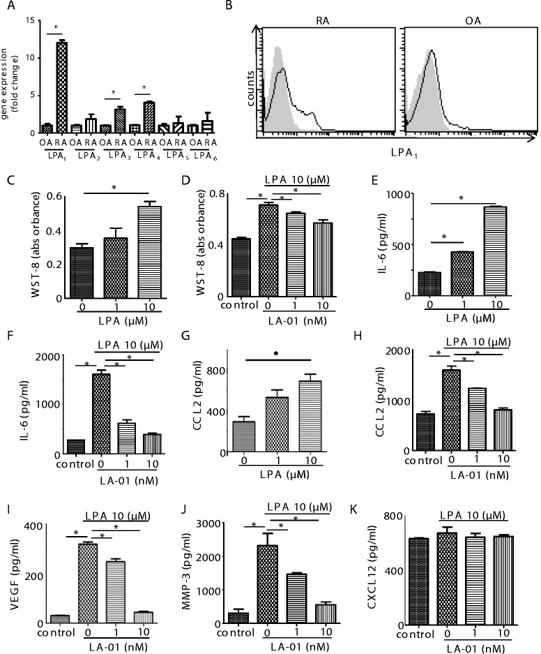
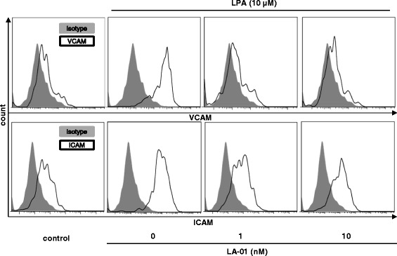
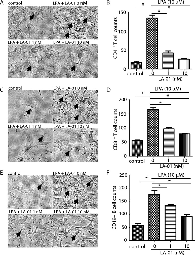
The expression of LPA1 mRNA and cell surface LPA1 was higher in RA FLSs than in FLSs from osteoarthritis tissue. Stimulation with LPA enhanced the proliferation of RA FLSs and the production of IL-6, VEGF, CCL2 and MMP-3 by FLSs, which were suppressed by an LPA1 inhibitor (LA-01). Ki16425, another LPA1 antagonist, also suppressed IL-6 production by LPA-stimulated RA FLSs. However, the production of CXCL12 was not altered by stimulation with LPA. LPA induced the pseudoemperipolesis of T and B cells cocultured with RA FLSs, which was suppressed by LPA1 inhibition. In addition, LPA enhanced the migration of RA FLSs and expression of vascular cell adhesion molecule and intercellular adhesion molecule on RA FLSs, which were also inhibited by an LPA1 antagonist.
Collectively, these results indicate that LPA-LPA1 signaling contributes to the activation of RA FLSs.
溶血磷脂酸(LPA)是一种可与G蛋白偶联受体(LPA1 - 6)结合的生物活性脂质。最近,我们报道LPA受体1(LPA1)的缺失改善了小鼠胶原诱导的关节炎,可能是通过抑制炎症细胞迁移、Th17分化和破骨细胞生成。在本研究中,我们检测了LPA - LPA1轴在类风湿关节炎(RA)患者滑膜组织来源的成纤维样滑膜细胞(FLS)的细胞增殖、细胞因子/趋化因子产生及淋巴细胞迁移中的重要性。
从RA患者的滑膜组织制备FLS。通过定量实时RT - PCR检测LPA1 - 6的表达。用流式细胞术分析细胞表面LPA1的表达。使用细胞计数试剂盒分析细胞增殖。通过酶联免疫吸附测定法检测白细胞介素6(IL - 6)、血管内皮生长因子(VEGF)、趋化因子(C - C基序)配体2(CCL2)、金属蛋白酶3(MMP - 3)和趋化因子(C - X - C基序)配体12(CXCL12)的产生。使用RA FLS与T或B细胞的共培养评估假包绕现象。通过划痕运动试验检测细胞运动性。用流式细胞术测定黏附分子的表达。
RA FLS中LPA1 mRNA和细胞表面LPA1的表达高于骨关节炎组织来源的FLS。LPA刺激增强了RA FLS的增殖以及FLS产生IL - 6、VEGF、CCL2和MMP - 3,这些均被LPA1抑制剂(LA - 01)所抑制。另一种LPA1拮抗剂Ki16425也抑制LPA刺激的RA FLS产生IL - 6。然而,LPA刺激未改变CXCL12的产生。LPA诱导与RA FLS共培养的T和B细胞的假包绕现象,这被LPA1抑制所抑制。此外,LPA增强了RA FLS的迁移以及RA FLS上血管细胞黏附分子和细胞间黏附分子的表达,这也被LPA1拮抗剂所抑制。
总体而言,这些结果表明LPA - LPA1信号传导有助于RA FLS的激活。