Klingner Christoph, Cherian Anoop V, Fels Johannes, Diesinger Philipp M, Aufschnaiter Roland, Maghelli Nicola, Keil Thomas, Beck Gisela, Tolić-Nørrelykke Iva M, Bathe Mark, Wedlich-Soldner Roland
Cellular Dynamics and Cell Patterning and Department of Molecular Structural Biology, Max Planck Institute of Biochemistry, 82152 Martinsried, Germany Institute of Cell Dynamics and Imaging and Cells-In-Motion Cluster of Excellence (EXC1003-CiM), University of Münster, 48149 Münster, Germany Institute of Cell Dynamics and Imaging and Cells-In-Motion Cluster of Excellence (EXC1003-CiM), University of Münster, 48149 Münster, Germany.
Cellular Dynamics and Cell Patterning and Department of Molecular Structural Biology, Max Planck Institute of Biochemistry, 82152 Martinsried, Germany.
J Cell Biol. 2014 Oct 13;207(1):107-21. doi: 10.1083/jcb.201402037.
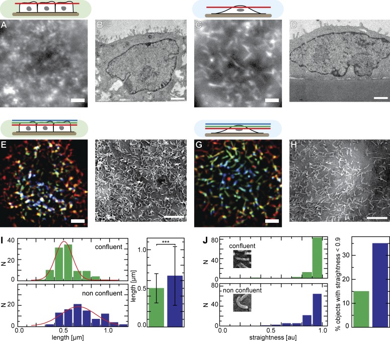
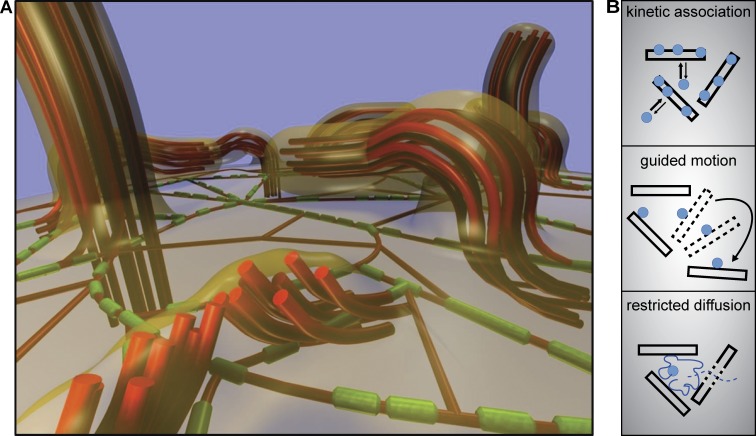
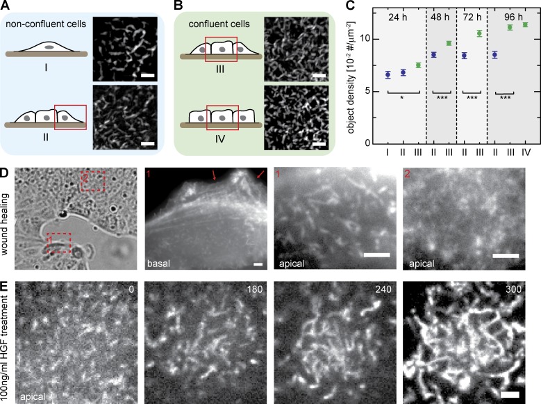
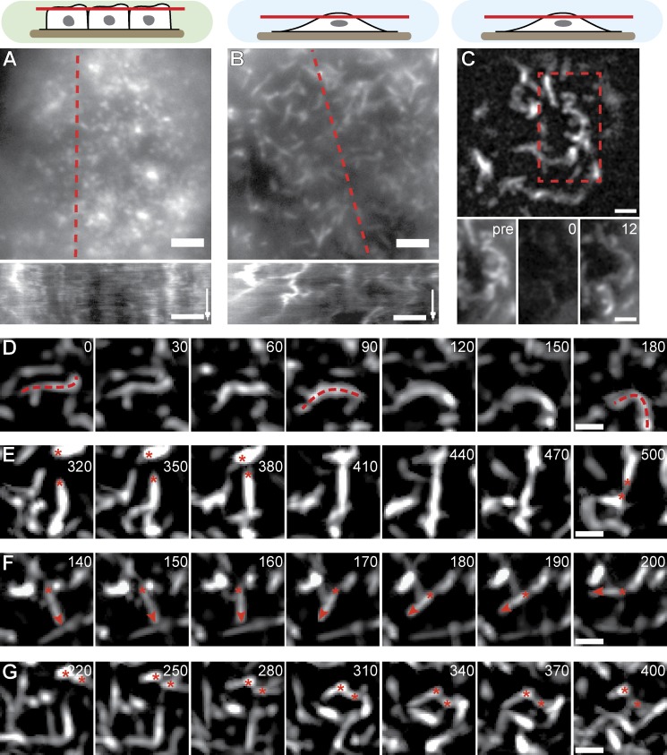
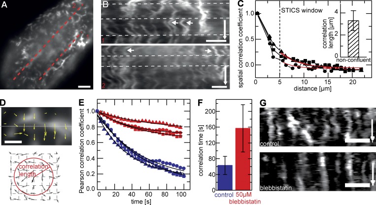
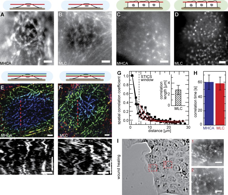
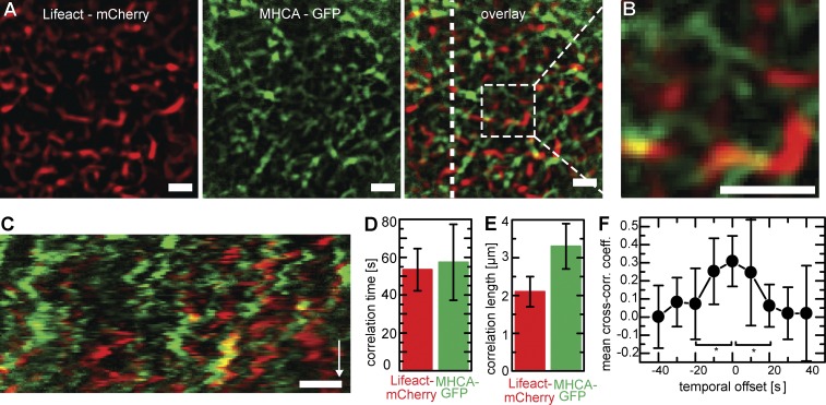
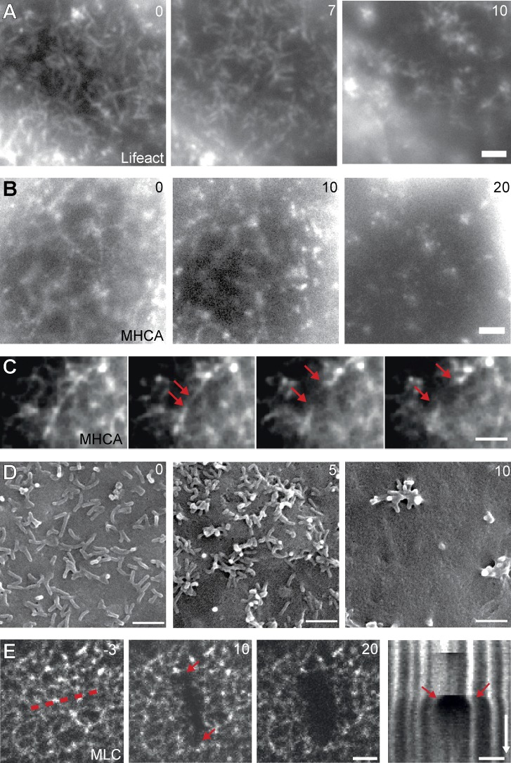
Although cortical actin plays an important role in cellular mechanics and morphogenesis, there is surprisingly little information on cortex organization at the apical surface of cells. In this paper, we characterize organization and dynamics of microvilli (MV) and a previously unappreciated actomyosin network at the apical surface of Madin-Darby canine kidney cells. In contrast to short and static MV in confluent cells, the apical surfaces of nonconfluent epithelial cells (ECs) form highly dynamic protrusions, which are often oriented along the plane of the membrane. These dynamic MV exhibit complex and spatially correlated reorganization, which is dependent on myosin II activity. Surprisingly, myosin II is organized into an extensive network of filaments spanning the entire apical membrane in nonconfluent ECs. Dynamic MV, myosin filaments, and their associated actin filaments form an interconnected, prestressed network. Interestingly, this network regulates lateral mobility of apical membrane probes such as integrins or epidermal growth factor receptors, suggesting that coordinated actomyosin dynamics contributes to apical cell membrane organization.
尽管皮质肌动蛋白在细胞力学和形态发生中起着重要作用,但令人惊讶的是,关于细胞顶端表面皮质组织的信息却很少。在本文中,我们描述了Madin-Darby犬肾细胞顶端表面微绒毛(MV)和一个以前未被重视的肌动球蛋白网络的组织和动态变化。与汇合细胞中短而静态的MV不同,未汇合上皮细胞(ECs)的顶端表面形成高度动态的突起,这些突起通常沿膜平面定向。这些动态MV表现出复杂且空间相关的重组,这依赖于肌球蛋白II的活性。令人惊讶的是,在未汇合的ECs中,肌球蛋白II被组织成一个跨越整个顶端膜的广泛细丝网络。动态MV、肌球蛋白细丝及其相关的肌动蛋白细丝形成一个相互连接的预应力网络。有趣的是,这个网络调节顶端膜探针(如整合素或表皮生长因子受体)的侧向移动性,表明协调的肌动球蛋白动态变化有助于顶端细胞膜的组织。