Hata Masayuki, Miyamoto Kazuaki, Oishi Akio, Makiyama Yukiko, Gotoh Norimoto, Kimura Yugo, Akagi Tadamichi, Yoshimura Nagahisa
Department of Ophthalmology and Visual Sciences, Kyoto University Graduate School of Medicine, Kyoto, Japan.
PLoS One. 2014 Nov 6;9(11):e112403. doi: 10.1371/journal.pone.0112403. eCollection 2014.
To compare the optic nerve head (ONH) structure between compressive optic neuropathy (CON) and glaucomatous optic neuropathy (GON), and to determine whether selected ONH quantitative parameters effectively discriminate between GON and CON, especially CON cases presenting with a glaucoma-like disc.
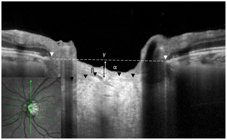
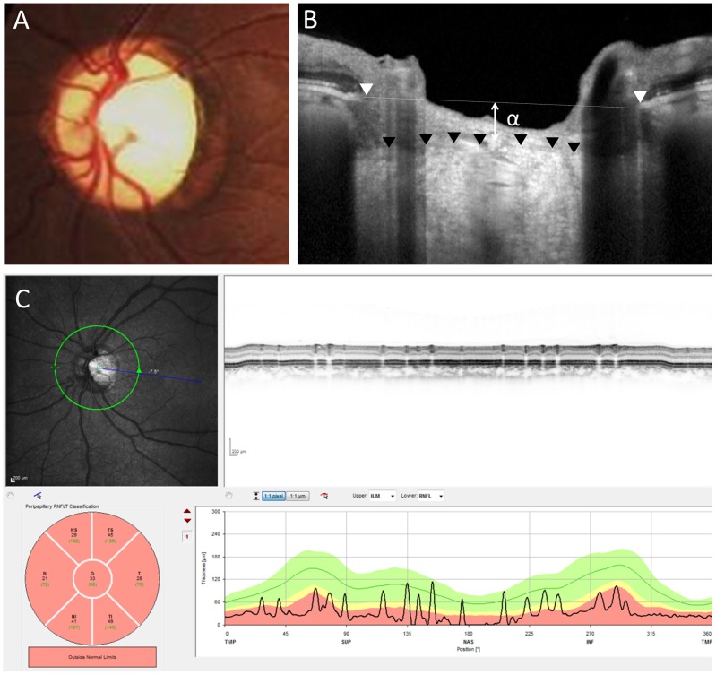
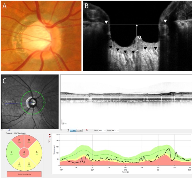
We prospectively assessed 34 patients with CON, 34 age-matched patients with moderate or severe GON, and 34 age-matched healthy control subjects. The quantitative parameters of ONH structure were compared using the Heidelberg Retina Tomograph 2 (HRT2) and Spectralis optical coherence tomography with an enhanced depth imaging method.
The mean and maximum cup depths of CON were significantly smaller than those with GON (P < 0.001 and P < 0.001, respectively). The distance between Bruch's membrane opening and anterior surface of the lamina cribrosa (BMO-anterior LC) of CON was also significantly smaller than that of glaucoma but was similar to that of the healthy group (P < 0.001 and P = 0.47, respectively). Based on Moorfields regression analysis of the glaucoma classification of HRT2, 15 eyes with CON were classified with a glaucoma-like disc. The cup/disc area ratio did not differ between cases of CON with a glaucoma-like disc and cases of GON (P = 0.16), but the BMO-anterior LC and mean and maximum cup depths of CON cases with a glaucoma-like disc were smaller than those in GON (P = 0.005, P = 0.003, and P = 0.001, respectively).
Measurements of the cup depths and the LC depth had good ability to differentiate between CON with a glaucoma-like disc and glaucoma. There was no laminar remodeling detected by laminar surface position in the patients with CON compared to those with GON.
比较压迫性视神经病变(CON)和青光眼性视神经病变(GON)之间的视神经乳头(ONH)结构,并确定选定的ONH定量参数是否能有效区分GON和CON,特别是呈现青光眼样视盘的CON病例。
我们前瞻性评估了34例CON患者、34例年龄匹配的中度或重度GON患者以及34例年龄匹配的健康对照者。使用海德堡视网膜断层扫描仪2(HRT2)和采用增强深度成像方法的Spectralis光学相干断层扫描比较ONH结构的定量参数。
CON的平均杯深和最大杯深显著小于GON(分别为P < 0.001和P < 0.001)。CON的布鲁赫膜开口与筛板前表面之间的距离(BMO-前LC)也显著小于青光眼患者,但与健康组相似(分别为P < 0.001和P = 0.47)。基于HRT2青光眼分类的穆尔菲尔德回归分析,15只CON眼被分类为青光眼样视盘。呈现青光眼样视盘的CON病例与GON病例之间的杯/盘面积比无差异(P = 0.16),但呈现青光眼样视盘的CON病例的BMO-前LC以及平均杯深和最大杯深均小于GON(分别为P = 0.005、P = 0.003和P = 0.001)。
杯深和筛板深度测量对区分呈现青光眼样视盘的CON和青光眼具有良好能力。与GON患者相比,未在CON患者中通过筛板表面位置检测到层状重塑。