• 文献检索
  • 文档翻译
  • 深度研究
  • 学术资讯
  • Suppr Zotero 插件Zotero 插件
  • 邀请有礼
  • 套餐&价格
  • 历史记录
应用&插件
Suppr Zotero 插件Zotero 插件浏览器插件Mac 客户端Windows 客户端微信小程序
定价
高级版会员购买积分包购买API积分包
服务
文献检索文档翻译深度研究API 文档MCP 服务
关于我们
关于 Suppr公司介绍联系我们用户协议隐私条款
关注我们

Suppr 超能文献

核心技术专利:CN118964589B侵权必究
粤ICP备2023148730 号-1Suppr @ 2026

文献检索

告别复杂PubMed语法,用中文像聊天一样搜索,搜遍4000万医学文献。AI智能推荐,让科研检索更轻松。

立即免费搜索

文件翻译

保留排版,准确专业,支持PDF/Word/PPT等文件格式,支持 12+语言互译。

免费翻译文档

深度研究

AI帮你快速写综述,25分钟生成高质量综述,智能提取关键信息,辅助科研写作。

立即免费体验

一种基于水平集分割和ReliefF特征选择的动态对比增强磁共振图像计算机辅助诊断系统。

A computer-aided diagnosis system for dynamic contrast-enhanced MR images based on level set segmentation and ReliefF feature selection.

作者信息

Pang Zhiyong, Zhu Dongmei, Chen Dihu, Li Li, Shao Yuanzhi

机构信息

School of Physics and Engineering, Sun Yat-sen University, Guangzhou 510275, China.

Imaging Diagnosis and Interventional Center, Cancer Center, The Sun Yat-sen University, Guangzhou 510060, China.

出版信息

Comput Math Methods Med. 2015;2015:450531. doi: 10.1155/2015/450531. Epub 2015 Jan 6.

DOI:10.1155/2015/450531
PMID:25628755
原文链接:https://pmc.ncbi.nlm.nih.gov/articles/PMC4300094/
Abstract

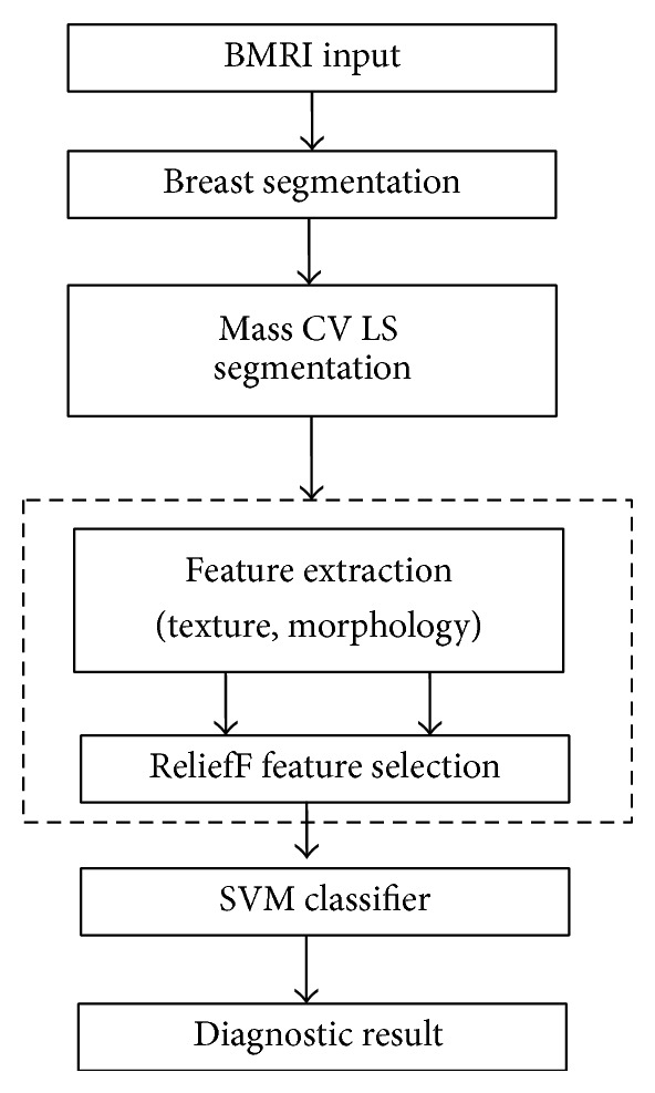
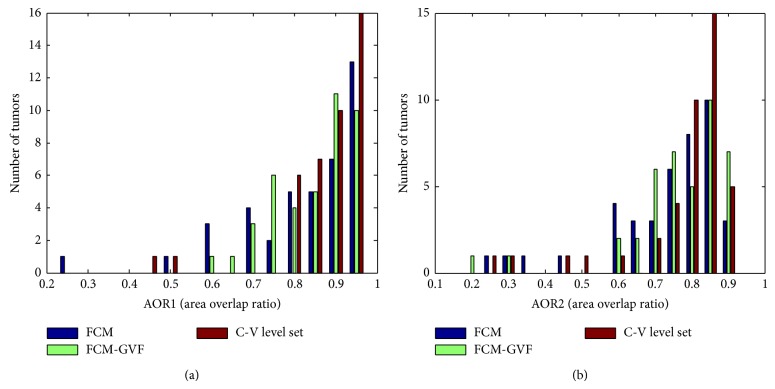
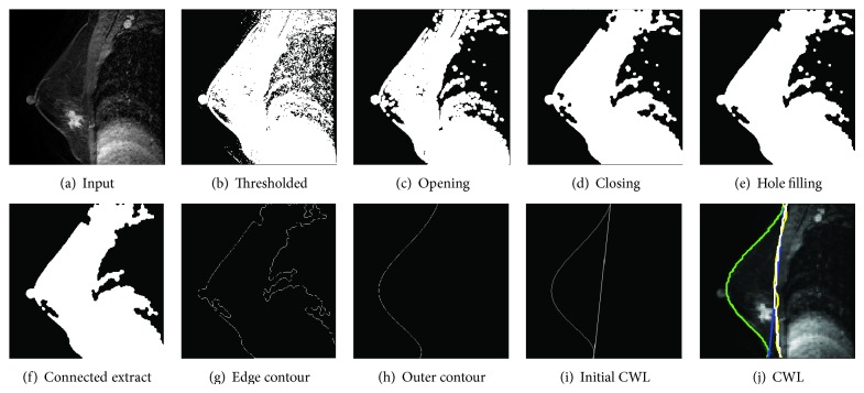
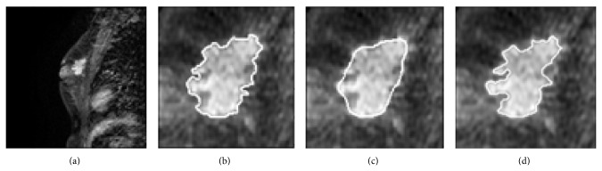
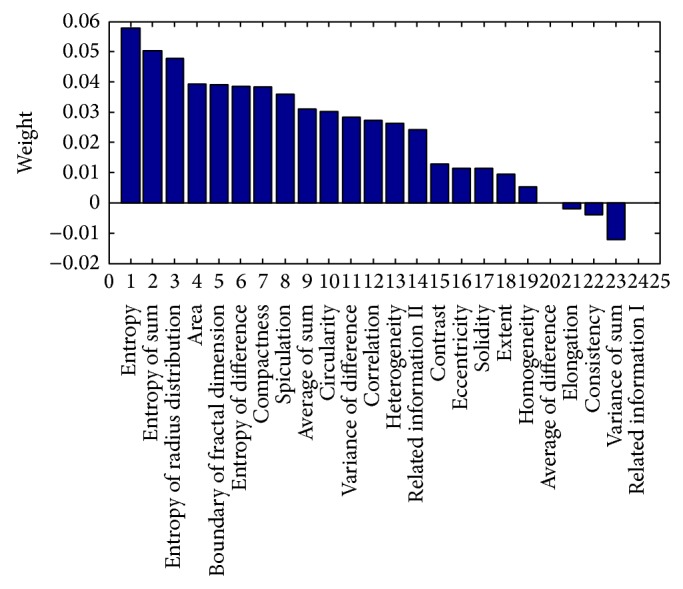
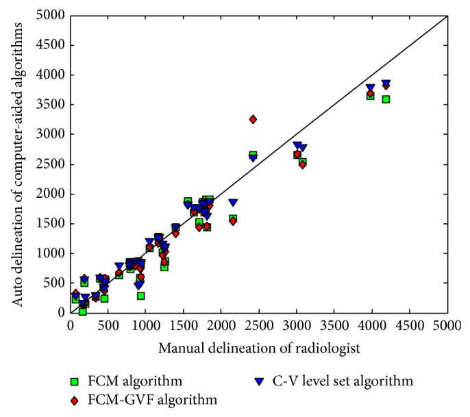
This study established a fully automated computer-aided diagnosis (CAD) system for the classification of malignant and benign masses via breast magnetic resonance imaging (BMRI). A breast segmentation method consisting of a preprocessing step to identify the air-breast interfacing boundary and curve fitting for chest wall line (CWL) segmentation was included in the proposed CAD system. The Chan-Vese (CV) model level set (LS) segmentation method was adopted to segment breast mass and demonstrated sufficiently good segmentation performance. The support vector machine (SVM) classifier with ReliefF feature selection was used to merge the extracted morphological and texture features into a classification score. The accuracy, sensitivity, and specificity measurements for the leave-half-case-out resampling method were 92.3%, 98.2%, and 76.2%, respectively. For the leave-one-case-out resampling method, the measurements were 90.0%, 98.7%, and 73.8%, respectively.

摘要

本研究建立了一种通过乳腺磁共振成像(BMRI)对恶性和良性肿块进行分类的全自动计算机辅助诊断(CAD)系统。所提出的CAD系统包括一种乳腺分割方法,该方法由识别空气-乳腺界面边界的预处理步骤和用于胸壁线(CWL)分割的曲线拟合组成。采用Chan-Vese(CV)模型水平集(LS)分割方法对乳腺肿块进行分割,并表现出足够好的分割性能。使用具有ReliefF特征选择的支持向量机(SVM)分类器将提取的形态学和纹理特征合并为一个分类分数。留半法重采样方法的准确度、灵敏度和特异性测量值分别为92.3%、98.2%和76.2%。对于留一法重采样方法,测量值分别为90.0%、98.7%和73.8%。

https://cdn.ncbi.nlm.nih.gov/pmc/blobs/ce5a/4300094/e5d1a8e55cb4/CMMM2015-450531.006.jpg
https://cdn.ncbi.nlm.nih.gov/pmc/blobs/ce5a/4300094/516fbc1a25dd/CMMM2015-450531.001.jpg
https://cdn.ncbi.nlm.nih.gov/pmc/blobs/ce5a/4300094/b25438045122/CMMM2015-450531.002.jpg
https://cdn.ncbi.nlm.nih.gov/pmc/blobs/ce5a/4300094/b39fe1f26ec7/CMMM2015-450531.003.jpg
https://cdn.ncbi.nlm.nih.gov/pmc/blobs/ce5a/4300094/f8654dfb07fa/CMMM2015-450531.004.jpg
https://cdn.ncbi.nlm.nih.gov/pmc/blobs/ce5a/4300094/65c386b8d76b/CMMM2015-450531.005.jpg
https://cdn.ncbi.nlm.nih.gov/pmc/blobs/ce5a/4300094/e5d1a8e55cb4/CMMM2015-450531.006.jpg

相似文献

1
A computer-aided diagnosis system for dynamic contrast-enhanced MR images based on level set segmentation and ReliefF feature selection.一种基于水平集分割和ReliefF特征选择的动态对比增强磁共振图像计算机辅助诊断系统。
Comput Math Methods Med. 2015;2015:450531. doi: 10.1155/2015/450531. Epub 2015 Jan 6.
2
Multilevel analysis of spatiotemporal association features for differentiation of tumor enhancement patterns in breast DCE-MRI.基于多时相分析的乳腺 DCE-MRI 肿瘤增强模式鉴别特征研究。
Med Phys. 2010 Aug;37(8):3940-56. doi: 10.1118/1.3446799.
3
Improving the Accuracy of Computer-aided Diagnosis for Breast MR Imaging by Differentiating between Mass and Nonmass Lesions.通过区分肿块和非肿块病变来提高乳腺磁共振成像计算机辅助诊断的准确性。
Radiology. 2016 Mar;278(3):679-88. doi: 10.1148/radiol.2015150241. Epub 2015 Sep 18.
4
Computer-aided diagnosis for dynamic contrast-enhanced breast MRI of mass-like lesions using a multiparametric model combining a selection of morphological, kinetic, and spatiotemporal features.基于形态学、动力学和时空特征选择的多参数模型对乳腺肿块样病变动态增强 MRI 的计算机辅助诊断。
Med Phys. 2012 Apr;39(4):1704-15. doi: 10.1118/1.3691178.
5
Computer-aided diagnosis of contrast-enhanced spectral mammography: A feasibility study.计算机辅助诊断对比增强光谱乳腺摄影:一项可行性研究。
Eur J Radiol. 2018 Jan;98:207-213. doi: 10.1016/j.ejrad.2017.11.024. Epub 2017 Dec 5.
6
A computer-aided diagnosis system for breast DCE-MRI at high spatiotemporal resolution.一种用于高时空分辨率乳腺动态对比增强磁共振成像的计算机辅助诊断系统。
Med Phys. 2016 Jan;43(1):84. doi: 10.1118/1.4937787.
7
Detection and classification of contrast-enhancing masses by a fully automatic computer-assisted diagnosis system for breast MRI.利用全自动计算机辅助诊断系统对乳腺 MRI 中的增强肿块进行检测和分类。
J Magn Reson Imaging. 2012 May;35(5):1077-88. doi: 10.1002/jmri.23516. Epub 2012 Jan 13.
8
Feature selection in computer-aided breast cancer diagnosis via dynamic contrast-enhanced magnetic resonance images.基于动态对比增强磁共振图像的计算机辅助乳腺癌诊断中的特征选择。
J Digit Imaging. 2013 Apr;26(2):198-208. doi: 10.1007/s10278-012-9506-2.
9
Characterization of spatiotemporal changes for the classification of dynamic contrast-enhanced magnetic-resonance breast lesions.动态对比增强磁共振乳腺病变分类的时空变化特征。
Artif Intell Med. 2013 Jun;58(2):101-14. doi: 10.1016/j.artmed.2013.03.002. Epub 2013 Mar 30.
10
Computer-aided characterization of mammographic masses: accuracy of mass segmentation and its effects on characterization.乳腺钼靶肿块的计算机辅助特征分析:肿块分割的准确性及其对特征分析的影响。
IEEE Trans Med Imaging. 2001 Dec;20(12):1275-84. doi: 10.1109/42.974922.

引用本文的文献

1
Use of a deep learning algorithm for non-mass enhancement on breast MRI: comparison with radiologists' interpretations at various levels.深度学习算法在乳腺 MRI 中非肿块样强化中的应用:与不同级别放射科医生解读的比较。
Jpn J Radiol. 2023 Oct;41(10):1094-1103. doi: 10.1007/s11604-023-01435-w. Epub 2023 Apr 18.
2
Infant birth weight estimation and low birth weight classification in United Arab Emirates using machine learning algorithms.利用机器学习算法估算阿联酋的婴儿出生体重和低出生体重分类。
Sci Rep. 2022 Jul 15;12(1):12110. doi: 10.1038/s41598-022-14393-6.
3
Intelligent Diagnostic Prediction and Classification Models for Detection of Kidney Disease.

本文引用的文献

1
Diagnosis of breast masses from dynamic contrast-enhanced and diffusion-weighted MR: a machine learning approach.基于动态对比增强和弥散加权磁共振的乳腺肿块诊断:一种机器学习方法。
PLoS One. 2014 Jan 31;9(1):e87387. doi: 10.1371/journal.pone.0087387. eCollection 2014.
2
A vector machine formulation with application to the computer-aided diagnosis of breast cancer from DCE-MRI screening examinations.一种应用于基于动态对比增强磁共振成像(DCE-MRI)筛查检查的乳腺癌计算机辅助诊断的向量机公式。
J Digit Imaging. 2014 Feb;27(1):145-51. doi: 10.1007/s10278-013-9621-8.
3
Automated chest wall line detection for whole-breast segmentation in sagittal breast MR images.
用于检测肾脏疾病的智能诊断预测和分类模型
Healthcare (Basel). 2022 Feb 14;10(2):371. doi: 10.3390/healthcare10020371.
4
A Deep Neural Network for Early Detection and Prediction of Chronic Kidney Disease.用于慢性肾脏病早期检测与预测的深度神经网络
Diagnostics (Basel). 2022 Jan 5;12(1):116. doi: 10.3390/diagnostics12010116.
5
Prediction of Clinical Outcome for High-Intensity Focused Ultrasound Ablation of Uterine Leiomyomas Using Multiparametric MRI Radiomics-Based Machine Leaning Model.基于多参数磁共振成像放射组学的机器学习模型预测高强度聚焦超声消融子宫肌瘤的临床结局
Front Oncol. 2021 Sep 10;11:618604. doi: 10.3389/fonc.2021.618604. eCollection 2021.
6
Deep Convolutional Neural Networks-Based Automatic Breast Segmentation and Mass Detection in DCE-MRI.基于深度卷积神经网络的动态对比增强磁共振成像中的乳腺自动分割与肿块检测
Comput Math Methods Med. 2020 May 5;2020:2413706. doi: 10.1155/2020/2413706. eCollection 2020.
7
Machine learning in breast MRI.乳腺磁共振成像中的机器学习
J Magn Reson Imaging. 2020 Oct;52(4):998-1018. doi: 10.1002/jmri.26852. Epub 2019 Jul 5.
8
Feature Selection for Object-Based Classification of High-Resolution Remote Sensing Images Based on the Combination of a Genetic Algorithm and Tabu Search.基于遗传算法和禁忌搜索的组合的高分辨率遥感图像基于对象分类的特征选择。
Comput Intell Neurosci. 2018 Jan 18;2018:6595792. doi: 10.1155/2018/6595792. eCollection 2018.
9
Construction of Metabolism Prediction Models for CYP450 3A4, 2D6, and 2C9 Based on Microsomal Metabolic Reaction System.基于微粒体代谢反应系统构建CYP450 3A4、2D6和2C9的代谢预测模型
Int J Mol Sci. 2016 Oct 9;17(10):1686. doi: 10.3390/ijms17101686.
10
Principles and methods for automatic and semi-automatic tissue segmentation in MRI data.MRI数据中自动和半自动组织分割的原理与方法
MAGMA. 2016 Apr;29(2):95-110. doi: 10.1007/s10334-015-0520-5. Epub 2016 Jan 11.
自动检测矢状位乳腺 MRI 图像中的胸壁线以进行全乳分割。
Med Phys. 2013 Apr;40(4):042301. doi: 10.1118/1.4793255.
4
Application of a spatially-weighted Relief algorithm for ranking genetic predictors of disease.基于空间加权 Relief 算法的疾病遗传预测因子排序应用
BioData Min. 2012 Dec 3;5(1):20. doi: 10.1186/1756-0381-5-20.
5
Computerized segmentation and characterization of breast lesions in dynamic contrast-enhanced MR images using fuzzy c-means clustering and snake algorithm.利用模糊 C 均值聚类和蛇算法对动态对比增强磁共振图像中的乳腺病变进行计算机分割和特征描述。
Comput Math Methods Med. 2012;2012:634907. doi: 10.1155/2012/634907. Epub 2012 Aug 21.
6
IMPST: A New Interactive Self-Training Approach to Segmentation Suspicious Lesions in Breast MRI.IMPST:一种用于乳腺MRI中可疑病变分割的新型交互式自我训练方法。
J Med Signals Sens. 2011 May;1(2):138-48.
7
Markov random field texture models.马尔可夫随机场纹理模型。
IEEE Trans Pattern Anal Mach Intell. 1983 Jan;5(1):25-39. doi: 10.1109/tpami.1983.4767341.
8
Beyond mammograms.
Sci Am. 2011 May;304(5):32, 34. doi: 10.1038/scientificamerican0511-32.
9
Characterization of masses in digital breast tomosynthesis: comparison of machine learning in projection views and reconstructed slices.数字乳腺断层合成中的肿块特征:在投影视图和重建切片中机器学习的比较。
Med Phys. 2010 Jul;37(7):3576-86. doi: 10.1118/1.3432570.
10
Treatment response assessment of breast masses on dynamic contrast-enhanced magnetic resonance scans using fuzzy c-means clustering and level set segmentation.使用模糊 C 均值聚类和水平集分割评估动态对比增强磁共振扫描中乳腺肿块的治疗反应。
Med Phys. 2009 Nov;36(11):5052-63. doi: 10.1118/1.3238101.