Raulf Alexandra, Horder Hannes, Tarnawski Laura, Geisen Caroline, Ottersbach Annika, Röll Wilhelm, Jovinge Stefan, Fleischmann Bernd K, Hesse Michael
Institute of Physiology I, Life and Brain Center, University of Bonn, Sigmund-Freud-Strasse 25, 53105, Bonn, Germany.
Basic Res Cardiol. 2015 May;110(3):33. doi: 10.1007/s00395-015-0489-2. Epub 2015 Apr 30.
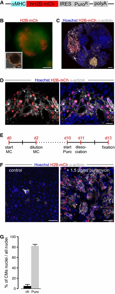
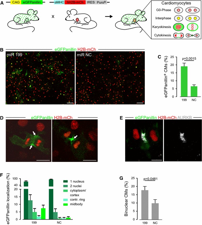
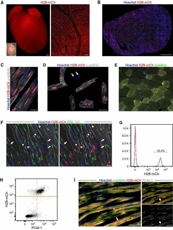
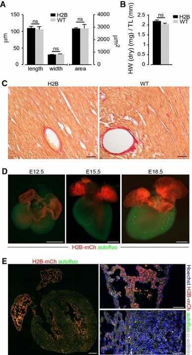
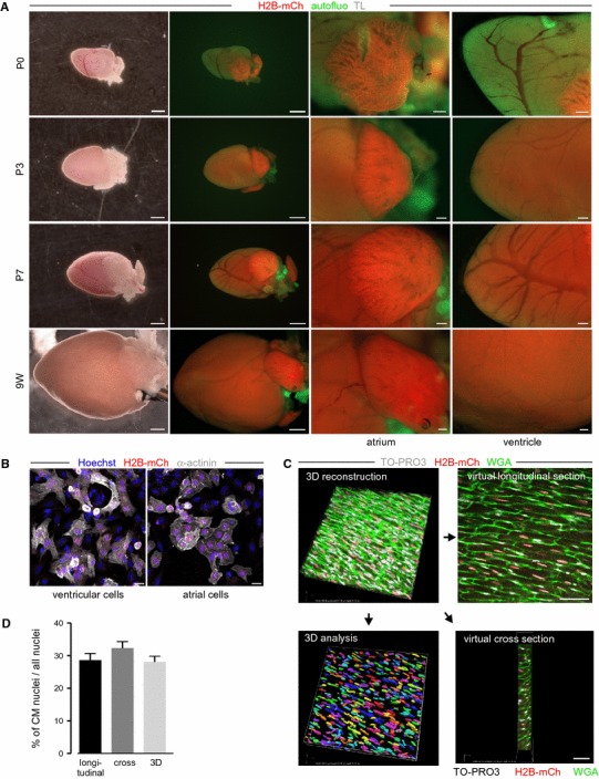
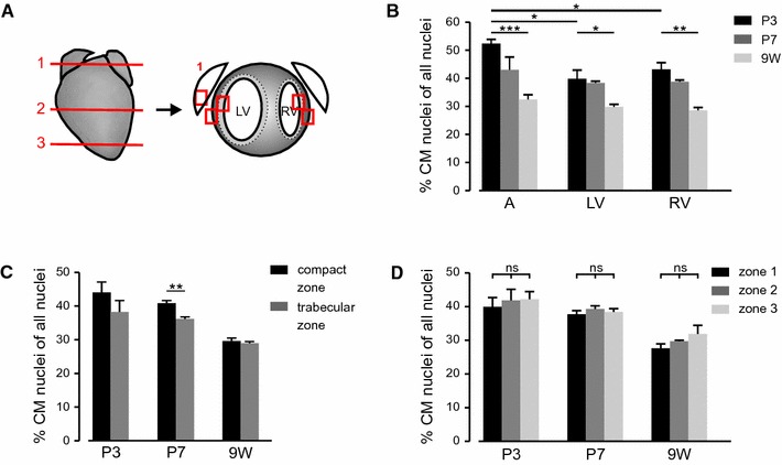
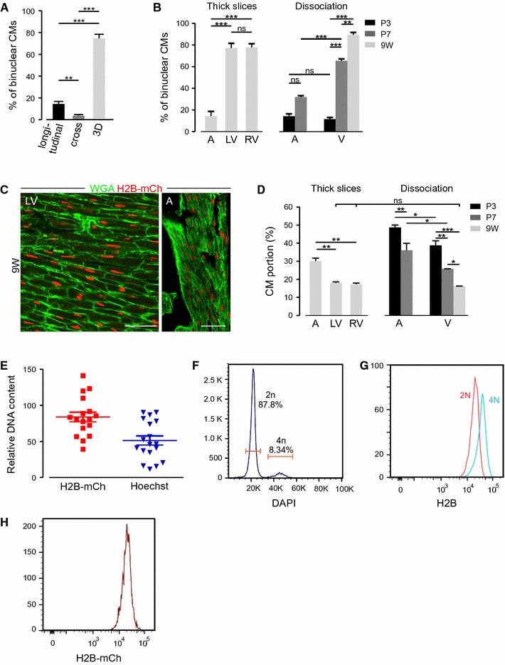
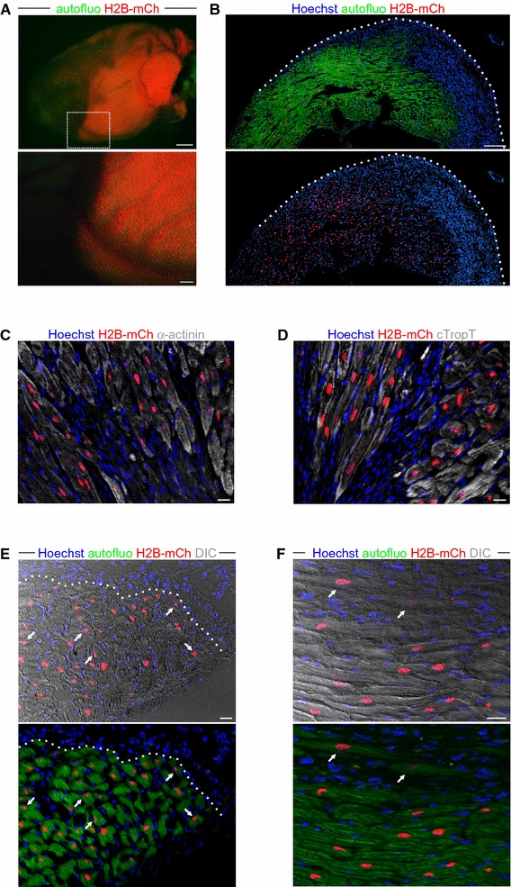
Even though the mammalian heart has been investigated for many years, there are still uncertainties in the fields of cardiac cell biology and regeneration with regard to exact fractions of cardiomyocytes (CMs) at different developmental stages, their plasticity after cardiac lesion and also their basal turnover rate. A main shortcoming is the accurate identification of CM and the demonstration of CM division. Therefore, an in vivo model taking advantage of a live reporter-based identification of CM nuclei and their cell cycle status is needed. In this technical report, we describe the generation and characterization of embryonic stem cells and transgenic mice expressing a fusion protein of human histone 2B and the red fluorescence protein mCherry under control of the CM specific αMHC promoter. This fluorescence label allows unequivocal identification and quantitation of CM nuclei and nuclearity in isolated cells and native tissue slices. In ventricles of adults, we determined a fraction of <20 % CMs and binucleation of 77-90 %, while in atria a CM fraction of 30 % and a binucleation index of 14 % were found. We combined this transgenic system with the CAG-eGFP-anillin transgene, which identifies cell division and established a novel screening assay for cell cycle-modifying substances in isolated, postnatal CMs. Our transgenic live reporter-based system enables reliable identification of CM nuclei and determination of CM fractions and nuclearity in heart tissue. In combination with CAG-eGFP-anillin-mice, the cell cycle status of CMs can be monitored in detail enabling screening for proliferation-inducing substances in vitro and in vivo.
尽管对哺乳动物心脏的研究已有多年,但在心脏细胞生物学和再生领域,关于不同发育阶段心肌细胞(CMs)的确切比例、心脏损伤后它们的可塑性以及它们的基础更新率仍存在不确定性。一个主要缺点是准确识别CMs并证明其分裂。因此,需要一种利用基于活细胞报告基因的CM细胞核及其细胞周期状态识别的体内模型。在本技术报告中,我们描述了在CM特异性αMHC启动子控制下表达人组蛋白2B与红色荧光蛋白mCherry融合蛋白的胚胎干细胞和转基因小鼠的产生及特性。这种荧光标记能够在分离的细胞和天然组织切片中明确识别和定量CM细胞核及核型。在成年心室中,我们确定CMs的比例小于20%,双核化比例为77 - 90%,而在心房中,CMs比例为30%,双核化指数为14%。我们将这个转基因系统与CAG - eGFP - anillin转基因相结合,该转基因可识别细胞分裂,并建立了一种用于筛选分离的出生后CMs中细胞周期修饰物质的新型检测方法。我们基于转基因活细胞报告基因的系统能够可靠地识别心脏组织中的CM细胞核,并确定CMs的比例和核型。与CAG - eGFP - anillin小鼠相结合,可以详细监测CMs的细胞周期状态,从而能够在体外和体内筛选增殖诱导物质。