Crocini C, Ferrantini C, Scardigli M, Coppini R, Mazzoni L, Lazzeri E, Pioner J M, Scellini B, Guo A, Song L S, Yan P, Loew L M, Tardiff J, Tesi C, Vanzi F, Cerbai E, Pavone F S, Sacconi L, Poggesi C
European Laboratory for Non-Linear Spectroscopy, 50019 Florence, Italy.
Division of Physiology, Department of Experimental and Clinical Medicine, University of Florence, 50134 Florence, Italy.
J Mol Cell Cardiol. 2016 Feb;91:42-51. doi: 10.1016/j.yjmcc.2015.12.013. Epub 2015 Dec 20.
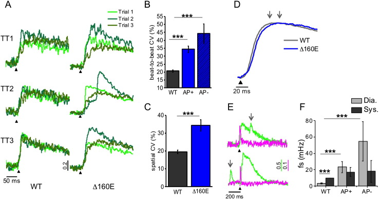
Abnormalities of cardiomyocyte Ca(2+) homeostasis and excitation-contraction (E-C) coupling are early events in the pathogenesis of hypertrophic cardiomyopathy (HCM) and concomitant determinants of the diastolic dysfunction and arrhythmias typical of the disease. T-tubule remodelling has been reported to occur in HCM but little is known about its role in the E-C coupling alterations of HCM. Here, the role of T-tubule remodelling in the electro-mechanical dysfunction associated to HCM is investigated in the Δ160E cTnT mouse model that expresses a clinically-relevant HCM mutation. Contractile function of intact ventricular trabeculae is assessed in Δ160E mice and wild-type siblings. As compared with wild-type, Δ160E trabeculae show prolonged kinetics of force development and relaxation, blunted force-frequency response with reduced active tension at high stimulation frequency, and increased occurrence of spontaneous contractions. Consistently, prolonged Ca(2+) transient in terms of rise and duration are also observed in Δ160E trabeculae and isolated cardiomyocytes. Confocal imaging in cells isolated from Δ160E mice reveals significant, though modest, remodelling of T-tubular architecture. A two-photon random access microscope is employed to dissect the spatio-temporal relationship between T-tubular electrical activity and local Ca(2+) release in isolated cardiomyocytes. In Δ160E cardiomyocytes, a significant number of T-tubules (>20%) fails to propagate action potentials, with consequent delay of local Ca(2+) release. At variance with wild-type, we also observe significantly increased variability of local Ca(2+) transient rise as well as higher Ca(2+)-spark frequency. Although T-tubule structural remodelling in Δ160E myocytes is modest, T-tubule functional defects determine non-homogeneous Ca(2+) release and delayed myofilament activation that significantly contribute to mechanical dysfunction.
心肌细胞钙(Ca2+)稳态异常和兴奋-收缩(E-C)偶联异常是肥厚型心肌病(HCM)发病机制中的早期事件,也是该疾病典型舒张功能障碍和心律失常的伴随决定因素。据报道,HCM中会发生T小管重塑,但其在HCM的E-C偶联改变中的作用尚不清楚。在此,我们在表达临床相关HCM突变的Δ160E cTnT小鼠模型中研究了T小管重塑在与HCM相关的电-机械功能障碍中的作用。评估了Δ160E小鼠和野生型同窝小鼠完整心室小梁的收缩功能。与野生型相比,Δ160E小梁显示出力量发展和松弛的动力学延长、在高刺激频率下力量-频率反应减弱且主动张力降低,以及自发收缩发生率增加。一致地,在Δ160E小梁和分离的心肌细胞中也观察到Ca2+瞬变在上升和持续时间方面延长。对从Δ160E小鼠分离的细胞进行共聚焦成像显示T小管结构有显著但适度的重塑。使用双光子随机存取显微镜剖析分离的心肌细胞中T小管电活动与局部Ca2+释放之间的时空关系。在Δ160E心肌细胞中,大量T小管(>20%)无法传播动作电位,导致局部Ca2+释放延迟。与野生型不同,我们还观察到局部Ca2+瞬变上升的变异性显著增加以及Ca2+火花频率更高。尽管Δ160E心肌细胞中的T小管结构重塑适度,但T小管功能缺陷决定了不均匀的Ca2+释放和延迟的肌丝激活,这显著导致了机械功能障碍。